WB
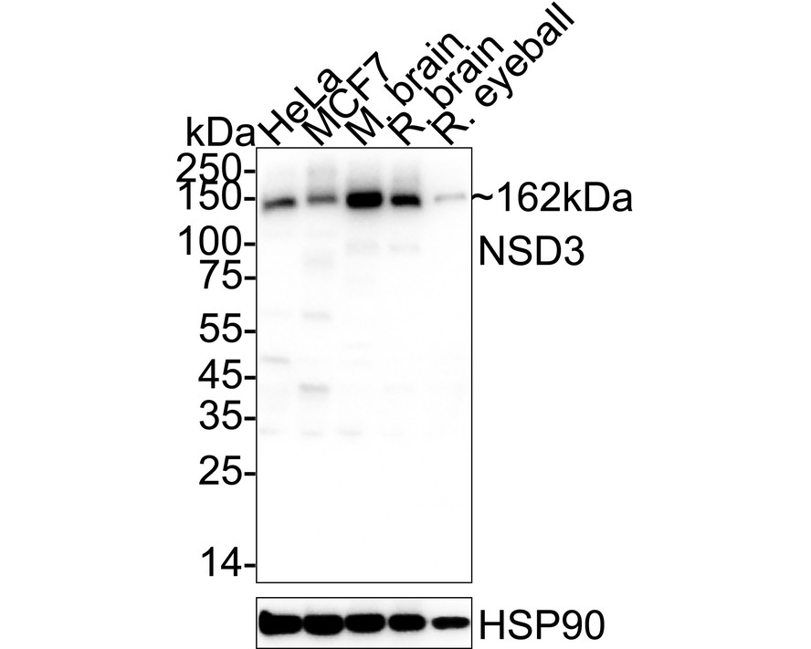
Western blot analysis of NSD3 on different lysates with Rabbit anti-NSD3 antibody at 1/10,000 dilution. Lane 1: HeLa cell lysate, Lane 2: MCF7 cell lysate, Lane 3: Mouse brain tissue lysate, Lane 4: Rat brain tissue lysate, Lane 5: Rat eyeball tissue lysate, Lysates/proteins at 20 µg/Lane. Exposure time: 10 seconds; 4-20% SDS-PAGE gel. Proteins were transferred to a PVDF membrane and blocked with 5% NFDM/TBST for 1 hour at room temperature. The primary antibody at 1/10,000 dilution was used in 5% NFDM/TBST at 4℃ overnight. Goat Anti-Rabbit IgG - HRP Secondary Antibody at 1/50,000 dilution was used for 1 hour at room temperature.IHC
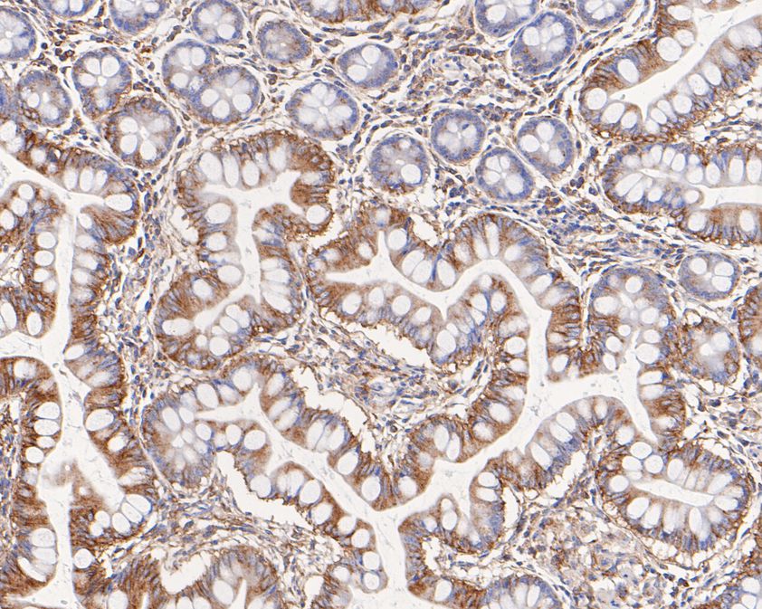
Immunohistochemical analysis of paraffin-embedded human small intestine tissue using anti-NSD3 antibody. The section was pre-treated using heat mediated antigen retrieval with Tris-EDTA buffer (pH 8.0-8.4) for 20 minutes.The tissues were blocked in 5% BSA for 30 minutes at room temperature, washed with ddH2O and PBS, and then probed with the primary antibody (1/200) for 30 minutes at room temperature. The detection was performed using an HRP conjugated compact polymer system. DAB was used as the chromogen. Tissues were counterstained with hematoxylin and mounted with DPX.IHC
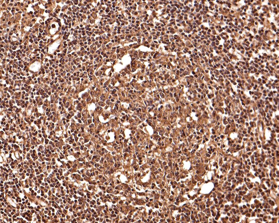
Immunohistochemical analysis of paraffin-embedded human lymph nodes tissue with Rabbit anti-NSD3 antibody at 1/400 dilution. The section was pre-treated using heat mediated antigen retrieval with Tris-EDTA buffer (pH 9.0) for 20 minutes. The tissues were blocked in 1% BSA for 20 minutes at room temperature, washed with ddH2O and PBS, and then probed with the primary antibody at 1/400 dilution for 1 hour at room temperature. The detection was performed using an HRP conjugated compact polymer system. DAB was used as the chromogen. Tissues were counterstained with hematoxylin and mounted with DPX.FC
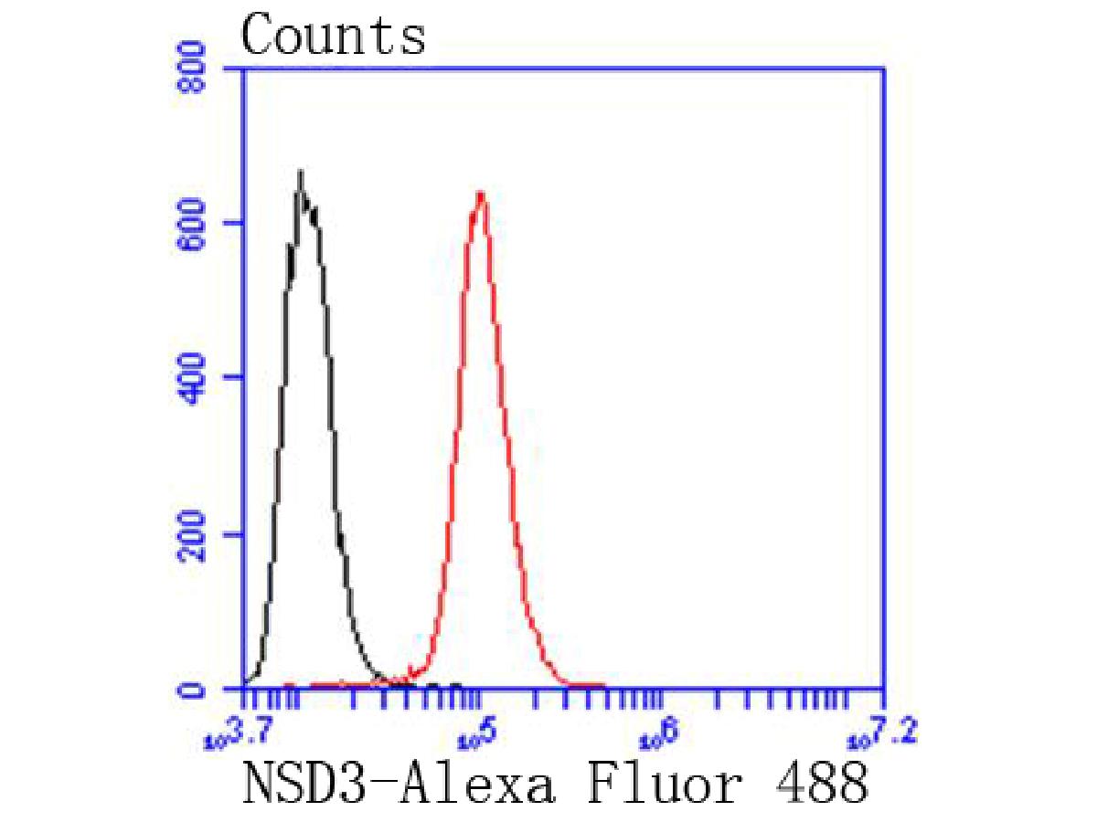
Flow cytometric analysis of NSD3 was done on Hela cells. The cells were fixed, permeabilized and stained with the primary antibody (1/50) (red). After incubation of the primary antibody at room temperature for an hour, the cells were stained with a Alexa Fluor 488-conjugated Goat anti-Rabbit IgG Secondary antibody at 1/1000 dilution for 30 minutes.Unlabelled sample was used as a control (cells without incubation with primary antibody; black).| Product Name | NSD3 Recombinant Rabbit Monoclonal Antibody |
|---|---|
| Antibody Type | Primary Antibodies |
| Product description | The deduced 1,437 amino acid NSD3 protein contains two PWWP domains involved in protein-protein interactions, five PHD-type zinc finger motifs found in chromatin-associated proteins, a SAC (SET-associated cys-rich) domain, a SET domain and a C-terminal C5HCH domain. Two NSD3 variants have been identified. The short variant comprised of 645 amino acids, arises from alternative polyadenylation and exon splicing and contains a single PWWP domain. A longer NSD3 variant, which is only expressed in HeLa cells, is comprised of 1,388 amino acid residues. The human WHSC1L1 gene, which encodes the NSD3 protein, shares 68% and 55% identity with mouse Nsd1 and human WHSC1, respectively. Highest expression of NSD3 is observed in brain, heart and skeletal muscle tissues; lower levels of NSD3 expression are observed in the liver and lungs. |
| Immunogen | Synthetic peptide within Human NSD3 aa 1-50 / 1,437. |
| Clonality | Monoclonal |
|---|---|
| Isotype | IgG |
| Host Species | Recombinant rabbit |
| Tested Applications | FCIHCWB |
| WB:1:10000 IHC:1:50-1:400 FC:1:50-1:100 |
|
| Species Reactivity | HumanMouseRat |
| Concentration | 1mg/ml |
| Purification | Protein A |
| Alternative Names | DKFZp667H044 antibody FLJ20353 antibody Histone lysine N methyltransferase NSD3 antibody Histone-lysine N-methyltransferase NSD3 antibody MGC126766 antibody MGC142029 antibody NSD 3 antibody NSD3_HUMAN antibody Nuclear set domain containing 3 antibody Nuclear SET domain containing protein 3 antibody Nuclear SET domain-containing protein 3 antibody pp14328 antibody Protein whistle antibody Whistle antibody WHSC1 like protein 1 antibody WHSC1-like 1 isoform 9 with methyltransferase activity to lysine antibody WHSC1-like protein 1 antibody WHSC1L1 antibody WHSC1L1 protein antibody Wolf Hirschhorn syndrome candidate 1 like 1 antibody Wolf-Hirschhorn syndrome candidate 1-like protein 1 antibody |
|---|---|
| Molecular Weight(MW) | 162 kDa |
| Cellular Localization | Nucleus, Chromosome. |
WB
Western blot analysis of NSD3 on different lysates with Rabbit anti-NSD3 antibody at 1/10,000 dilution. Lane 1: HeLa cell lysate, Lane 2: MCF7 cell lysate, Lane 3: Mouse brain tissue lysate, Lane 4: Rat brain tissue lysate, Lane 5: Rat eyeball tissue lysate, Lysates/proteins at 20 µg/Lane. Exposure time: 10 seconds; 4-20% SDS-PAGE gel. Proteins were transferred to a PVDF membrane and blocked with 5% NFDM/TBST for 1 hour at room temperature. The primary antibody at 1/10,000 dilution was used in 5% NFDM/TBST at 4℃ overnight. Goat Anti-Rabbit IgG - HRP Secondary Antibody at 1/50,000 dilution was used for 1 hour at room temperature.
IHC
Immunohistochemical analysis of paraffin-embedded human small intestine tissue using anti-NSD3 antibody. The section was pre-treated using heat mediated antigen retrieval with Tris-EDTA buffer (pH 8.0-8.4) for 20 minutes.The tissues were blocked in 5% BSA for 30 minutes at room temperature, washed with ddH2O and PBS, and then probed with the primary antibody (1/200) for 30 minutes at room temperature. The detection was performed using an HRP conjugated compact polymer system. DAB was used as the chromogen. Tissues were counterstained with hematoxylin and mounted with DPX.
IHC
Immunohistochemical analysis of paraffin-embedded human lymph nodes tissue with Rabbit anti-NSD3 antibody at 1/400 dilution. The section was pre-treated using heat mediated antigen retrieval with Tris-EDTA buffer (pH 9.0) for 20 minutes. The tissues were blocked in 1% BSA for 20 minutes at room temperature, washed with ddH2O and PBS, and then probed with the primary antibody at 1/400 dilution for 1 hour at room temperature. The detection was performed using an HRP conjugated compact polymer system. DAB was used as the chromogen. Tissues were counterstained with hematoxylin and mounted with DPX.
FC
Flow cytometric analysis of NSD3 was done on Hela cells. The cells were fixed, permeabilized and stained with the primary antibody (1/50) (red). After incubation of the primary antibody at room temperature for an hour, the cells were stained with a Alexa Fluor 488-conjugated Goat anti-Rabbit IgG Secondary antibody at 1/1000 dilution for 30 minutes.Unlabelled sample was used as a control (cells without incubation with primary antibody; black).| Application Notes | WB:1:10000 IHC:1:50-1:400 FC:1:50-1:100 |
|---|
| Form | Liquid |
|---|---|
| Storage Instructions | Store at +4℃ after thawing. Aliquot store at -20℃ or -80℃. Avoid repeated freeze / thaw cycles. |
| Storage Buffer | 1*TBS (pH7.4), 1%BSA, 40%Glycerol. Preservative: 0.05% Sodium Azide. |


