Application
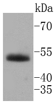
Fig1: Western blot analysis of HNF-4-alpha on human kidney lysates using anti-HNF-4-alpha antibody at 1/1,000 dilution.Application
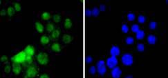
Fig2: ICC staining HNF-4-alpha in SW480 cells (green). The nuclear counter stain is DAPI (blue). Cells were fixed in paraformaldehyde, permeabilised with 0.25% Triton X100/PBS.Application
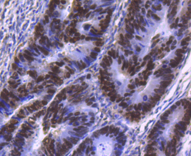
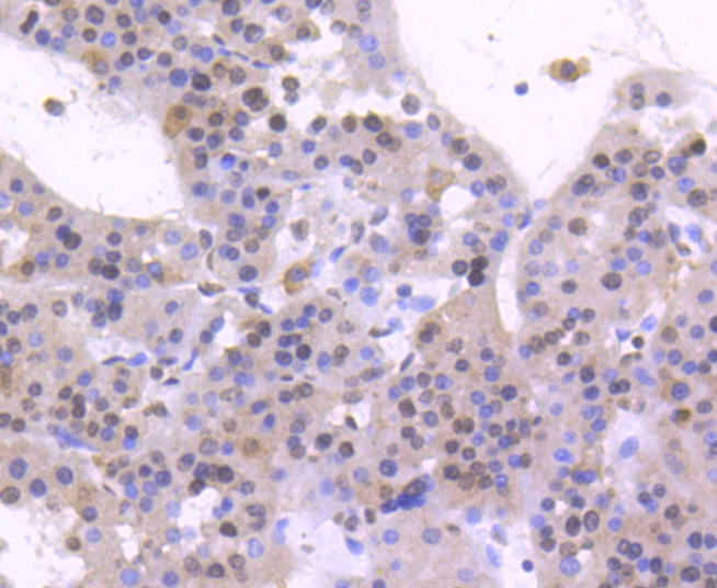
Fig3: Immunohistochemical analysis of paraffin-embedded human colon cancer tissue using anti-HNF-4-alpha antibody. Counter stained with hematoxylin.Application
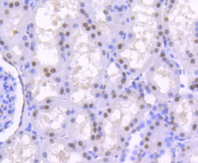
Fig4: Immunohistochemical analysis of paraffin-embedded human kidney tissue using anti-HNF-4-alpha antibody. Counter stained with hematoxylin.Application
Fig5: Immunohistochemical analysis of paraffin-embedded human liver cancer tissue using anti-HNF-4-alpha antibody. Counter stained with hematoxylin.Application
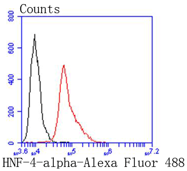
Fig6: Flow cytometric analysis of HepG2 cells with HNF-4-alpha antibody at 1/50 dilution (red) compared with an unlabelled control (cells without incubation with primary antibody; black). Alexa Fluor 488-conjugated goat anti rabbit IgG was used as the secondary antibody| Product Name | HNF-4-alpha [SN72-03] |
|---|---|
| Antibody Type | Primary Antibodies |
| Antigen Alias | FLJ39654 antibody FRTS4 antibody Hepatic nuclear factor 4 alpha antibody Hepatocyte nuclear factor 4 alpha antibody Hepatocyte nuclear factor 4 antibody Hepatocyte nuclear factor 4-alpha antibody HNF 4 alpha antibody HNF 4 antibody HNF-4-alpha antibody HNF4 antibody HNF4A antibody HNF4A_HUMAN antibody HNF4a7 antibody HNF4a8 antibody HNF4a9 antibody Hnf4alpha antibody HNF4alpha10/11/12 antibody MODY 1 antibody MODY antibody MODY1 antibody NR2A1 antibody NR2A21 antibody Nuclear receptor subfamily 2 group A member 1 antibody OTTHUMP00000031060 antibody OTTHUMP00000031062 antibody TCF 14 antibody TCF antibody TCF-14 antibody TCF14 antibody Tcf4 antibody Transcription factor 14, hepatic nuclear factor antibody Transcription factor 14 antibody Transcription factor HNF 4 antibody Transcription factor HNF-4 antibody Transcription factor HNF4 antibody |
| Product description | HNF-4 alpha is a transcription factor that binds DNA as a homodimer. HNF-4 alpha is important in liver, kidney, and intestinal development. It has also been intensely studied as one of a variety of genes responsible for diabetes mellitus. HNF-4 alpha has been shown in knock out mice to be essential for the morphogenic and functional differentiation of hepatocytes. HNF-4 alpha is a dominant regulator of epithelial phenotypes able to drive the mesenchymal-to-epithelial transition when expressed in fibroblasts. |
| Immunogen | recombinant protein |
| Clonality | Monoclonal |
|---|---|
| Isotype | IgG |
| Host Species | Recombinant rabbit |
| Tested Applications | WBICC/IFIHCFC |
| WB:1:1,000 ICC:1:50-1:200 IHC:1:100-1:500 FC:1:50-1:100 |
|
| Species Reactivity | Human |
| Concentration | 1mg/ml |
| Alternative Names | FLJ39654 antibody
FRTS4 antibody
Hepatic nuclear factor 4 alpha antibody
Hepatocyte nuclear factor 4 alpha antibody
Hepatocyte nuclear factor 4 antibody
Hepatocyte nuclear factor 4-alpha antibody
HNF 4 alpha antibody
HNF 4 antibody
HNF-4-alpha antibody
HNF4 antibody
HNF4A antibody
HNF4A_HUMAN antibody
HNF4a7 antibody
HNF4a8 antibody
HNF4a9 antibody
Hnf4alpha antibody
HNF4alpha10/11/12 antibody
MODY 1 antibody
MODY antibody
MODY1 antibody
NR2A1 antibody
NR2A21 antibody
Nuclear receptor subfamily 2 group A member 1 antibody
OTTHUMP00000031060 antibody
OTTHUMP00000031062 antibody
TCF 14 antibody
TCF antibody
TCF-14 antibody
TCF14 antibody
Tcf4 antibody
Transcription factor 14 hepatic nuclear factor antibody Transcription factor 14 antibody Transcription factor HNF 4 antibody Transcription factor HNF-4 antibody Transcription factor HNF4 antibody |
|---|---|
| Molecular Weight(MW) | 53 kDa |
| Cellular Localization | Nucleus. |
| SwissProt ID | P41235 |
|---|
Application
Fig1: Western blot analysis of HNF-4-alpha on human kidney lysates using anti-HNF-4-alpha antibody at 1/1,000 dilution.
Application
Fig2: ICC staining HNF-4-alpha in SW480 cells (green). The nuclear counter stain is DAPI (blue). Cells were fixed in paraformaldehyde, permeabilised with 0.25% Triton X100/PBS.
Application
Fig3: Immunohistochemical analysis of paraffin-embedded human colon cancer tissue using anti-HNF-4-alpha antibody. Counter stained with hematoxylin.
Application
Fig4: Immunohistochemical analysis of paraffin-embedded human kidney tissue using anti-HNF-4-alpha antibody. Counter stained with hematoxylin.
Application
Fig5: Immunohistochemical analysis of paraffin-embedded human liver cancer tissue using anti-HNF-4-alpha antibody. Counter stained with hematoxylin.
Application
Fig6: Flow cytometric analysis of HepG2 cells with HNF-4-alpha antibody at 1/50 dilution (red) compared with an unlabelled control (cells without incubation with primary antibody; black). Alexa Fluor 488-conjugated goat anti rabbit IgG was used as the secondary antibody| Positive Control | SW480, human colon cancer tissue, human kidney tissue, human liver cancer tissue. |
|---|---|
| Application Notes | WB:1:1,000 ICC:1:50-1:200 IHC:1:100-1:500 FC:1:50-1:100 |
| Form | Liquid |
|---|---|
| Storage Instructions | Store at +4℃ after thawing. Aliquot store at -20℃ or -80℃. Avoid repeated freeze / thaw cycles. |
| Storage Buffer | 1*TBS (pH7.4), 1%BSA, 40%Glycerol. Preservative: 0.05% Sodium Azide. |


