Application
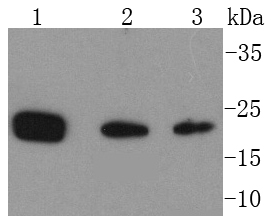
Fig1: Western blot analysis of PARK7 on different lysates using anti-PARK7 antibody at 1/1,000 dilution. Positive control: Lane 1: Hela Lane 2: Jurkat Lane 3: NIH/3T3Application
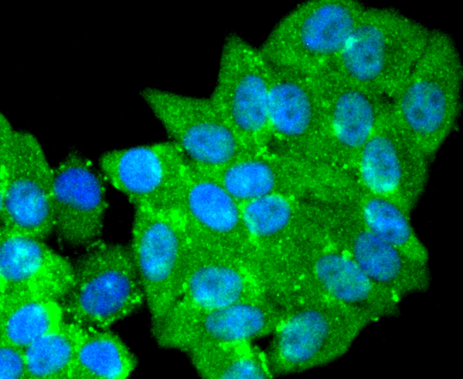
Fig2: ICC staining PARK7 in HepG2 cells (green). The nuclear counter stain is DAPI (blue). Cells were fixed in paraformaldehyde, permeabilised with 0.25% Triton X100/PBS.Application
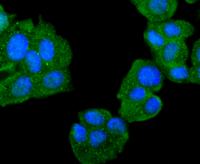
Fig3: ICC staining PARK7 in MCF-7 cells (green). The nuclear counter stain is DAPI (blue). Cells were fixed in paraformaldehyde, permeabilised with 0.25% Triton X100/PBS.Application
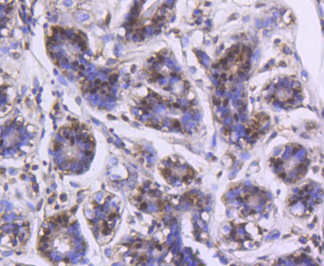
Fig4: Immunohistochemical analysis of paraffin-embedded human breast tissue using anti-PARK7 antibody. Counter stained with hematoxylin.Application
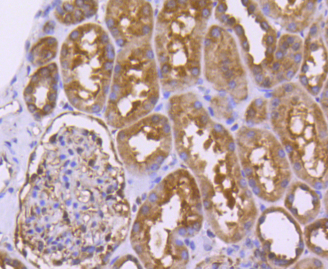
Fig5: Immunohistochemical analysis of paraffin-embedded human kidney tissue using anti-PARK7 antibody. Counter stained with hematoxylin.Application
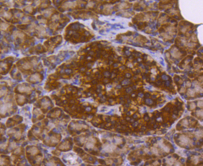
Fig6: Immunohistochemical analysis of paraffin-embedded human pancreas tissue using anti-PARK7 antibody. Counter stained with hematoxylin.Application
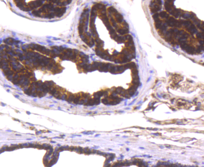
Fig7: Immunohistochemical analysis of paraffin-embedded mouse prostate tissue using anti-PARK7 antibody. Counter stained with hematoxylin.Application
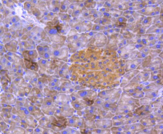
Fig8: Immunohistochemical analysis of paraffin-embedded mouse pancreas tissue using anti-PARK7 antibody. Counter stained with hematoxylin.Application
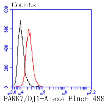
Fig9: Flow cytometric analysis of HepG2 cells with PARK7 antibody at 1/50 dilution (red) compared with an unlabelled control (cells without incubation with primary antibody; black). Alexa Fluor 488-conjugated goat anti rabbit IgG was used as the secondary antibody.| Product Name | PARK7/DJ1 [SN07-21] |
|---|---|
| Antibody Type | Primary Antibodies |
| Antigen Alias | CAP1 antibody DJ-1 antibody DJ1 antibody DJ1 protein antibody Epididymis secretory sperm binding protein Li 67p antibody FLJ27376 antibody FLJ34360 antibody FLJ92274 antibody HEL S 67p antibody Oncogene DJ1 antibody OTTHUMP00000001348 antibody OTTHUMP00000001349 antibody OTTHUMP00000001350 antibody OTTHUMP00000001351 antibody PARK7 antibody PARK7_HUMAN antibody Parkinson disease (autosomal recessive, early onset) 7 antibody Parkinson disease protein 7 antibody Parkinson protein 7 antibody Protein DJ-1 antibody SP22 antibody |
| Product description | PARK7/DJ1 is a ubiquitously expressed protein involved in various cellular processes including cell proliferation, RNA-binding, and oxidative stress. The protein has been found to colocalize within a subset of pathologic tau inclusions in a diverse group of neurodegenerative disorders known as tauopathies. Defects in PARK7/DJ1 are the cause of autosomal recessive early-onset Parkinson's disease 7 (PARK7). Parkinson's disease (PD) is a complex, multifactorial disorder that typically manifests after the age of 50 years. The disease is characterized by bradykinesia, resting tremor, muscular rigidity and postural instability. The pathology involves the loss of dopaminergic neurons in the substantia nigra and the presence of Lewy bodies (intraneuronal accumulations of aggregated proteins), in surviving neurons in various areas of the brain. PARK7 is characterized by onset before 40 years and slow progression. It has also been suggested that PARK7/DJ1 is a mitogen dependent oncogene product involved in Ras related signal transduction pathways. |
| Immunogen | recombinant protein |
| Clonality | Monoclonal |
|---|---|
| Isotype | IgG |
| Host Species | Recombinant rabbit |
| Tested Applications | WBICC/IFIHCIPFC |
| WB:1:1,000-1:2,000 ICC:1:100-1:500 IHC:1:100-1:500 FC:1:50-1:100 |
|
| Species Reactivity | HumanMouseRat |
| Concentration | 1mg/ml |
| Alternative Names | CAP1 antibody
DJ-1 antibody
DJ1 antibody
DJ1 protein antibody
Epididymis secretory sperm binding protein Li 67p antibody
FLJ27376 antibody
FLJ34360 antibody
FLJ92274 antibody
HEL S 67p antibody
Oncogene DJ1 antibody
OTTHUMP00000001348 antibody
OTTHUMP00000001349 antibody
OTTHUMP00000001350 antibody
OTTHUMP00000001351 antibody
PARK7 antibody
PARK7_HUMAN antibody
Parkinson disease (autosomal recessive early onset) 7 antibody Parkinson disease protein 7 antibody Parkinson protein 7 antibody Protein DJ-1 antibody SP22 antibody |
|---|---|
| Molecular Weight(MW) | 20 kDa |
| Cellular Localization | Cell membrane, Cytoplasm, Nucleus, Membrane raft, Mitochondrion. |
Application
Fig1: Western blot analysis of PARK7 on different lysates using anti-PARK7 antibody at 1/1,000 dilution. Positive control: Lane 1: Hela Lane 2: Jurkat Lane 3: NIH/3T3
Application
Fig2: ICC staining PARK7 in HepG2 cells (green). The nuclear counter stain is DAPI (blue). Cells were fixed in paraformaldehyde, permeabilised with 0.25% Triton X100/PBS.
Application
Fig3: ICC staining PARK7 in MCF-7 cells (green). The nuclear counter stain is DAPI (blue). Cells were fixed in paraformaldehyde, permeabilised with 0.25% Triton X100/PBS.
Application
Fig4: Immunohistochemical analysis of paraffin-embedded human breast tissue using anti-PARK7 antibody. Counter stained with hematoxylin.
Application
Fig5: Immunohistochemical analysis of paraffin-embedded human kidney tissue using anti-PARK7 antibody. Counter stained with hematoxylin.
Application
Fig6: Immunohistochemical analysis of paraffin-embedded human pancreas tissue using anti-PARK7 antibody. Counter stained with hematoxylin.
Application
Fig7: Immunohistochemical analysis of paraffin-embedded mouse prostate tissue using anti-PARK7 antibody. Counter stained with hematoxylin.
Application
Fig8: Immunohistochemical analysis of paraffin-embedded mouse pancreas tissue using anti-PARK7 antibody. Counter stained with hematoxylin.
Application
Fig9: Flow cytometric analysis of HepG2 cells with PARK7 antibody at 1/50 dilution (red) compared with an unlabelled control (cells without incubation with primary antibody; black). Alexa Fluor 488-conjugated goat anti rabbit IgG was used as the secondary antibody.| Positive Control | Jurkat, MCF-7, Hela, HepG2, NIH/3T3, human breast tissue, mouse pancreas tissue, human kidney tissue, human pancreas tissue, mouse prostate tissue. |
|---|---|
| Application Notes | WB:1:1,000-1:2,000 ICC:1:100-1:500 IHC:1:100-1:500 FC:1:50-1:100 |
| Form | Liquid |
|---|---|
| Storage Instructions | Store at +4℃ after thawing. Aliquot store at -20℃ or -80℃. Avoid repeated freeze / thaw cycles. |
| Storage Buffer | 1*TBS (pH7.4), 1%BSA, 40%Glycerol. Preservative: 0.05% Sodium Azide. |


