WB
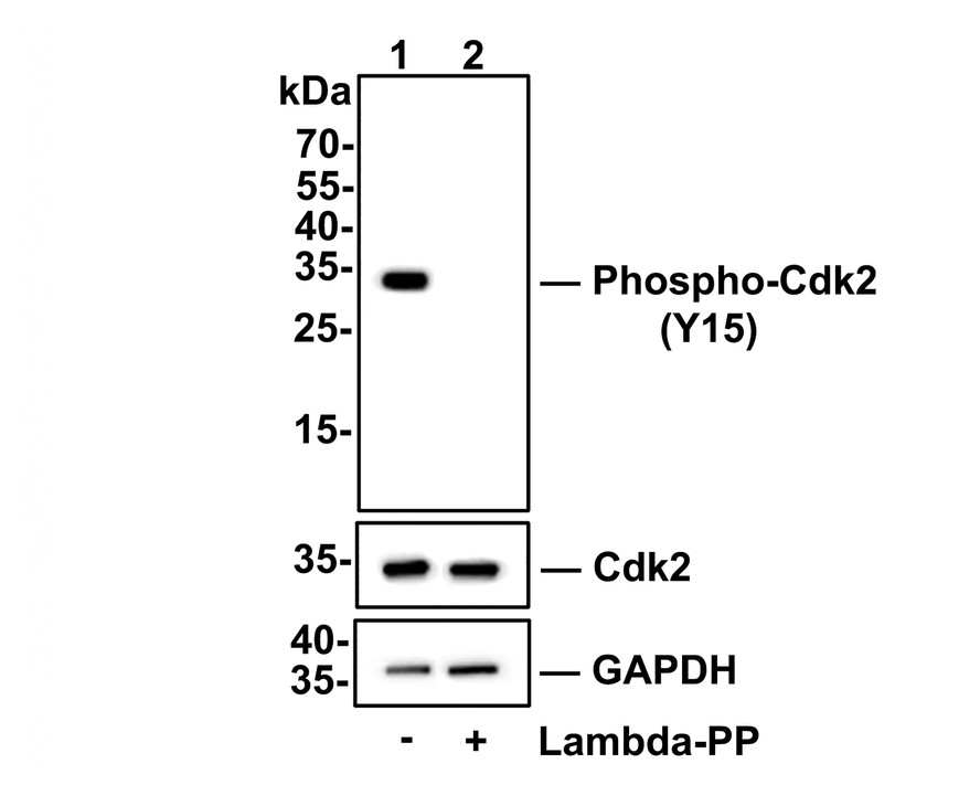
Western blot analysis of Phospho-Cdk2(Y15) on Hela cell lysates. Lane 1: Hela cells, whole cell lysate, 10ug/lane, Lane 2: Hela cells treated with 2.8ug/ul lambda-PP for 30 minutes, whole cell lysates, 10ug/lane, All lanes : Anti-Phospho-Cdk2(Y15) antibody at 1/500 dilution. Anti-Cdk2 antibody at 1/500 dilution. Anti-GAPDH antibody at 1/10,000 dilution. Goat Anti-Rabbit IgG H&L (HRP)at 1/200,000 dilution. Blocking and diluting buffer: 5% BSA. Exposure time: 30 seconds.IHC
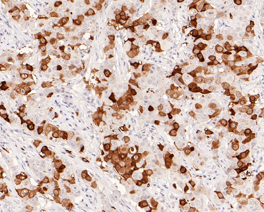
Immunohistochemical analysis of paraffin-embedded human breast carcinoma tissue with Rabbit anti-Phospho-Cdk2 (Y15) antibody at 1/1,000 dilution. The section was pre-treated using heat mediated antigen retrieval with sodium citrate buffer (pH 6.0) for 2 minutes. The tissues were blocked in 1% BSA for 20 minutes at room temperature, washed with ddH2O and PBS, and then probed with the primary antibody at 1/1,000 dilution for 1 hour at room temperature. The detection was performed using an HRP conjugated compact polymer system. DAB was used as the chromogen. Tissues were counterstained with hematoxylin and mounted with DPX.IHC
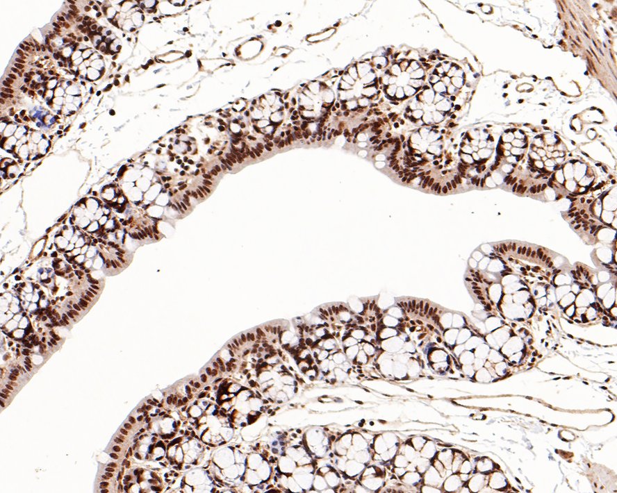
Immunohistochemical analysis of paraffin-embedded mouse small intestine tissue with Rabbit anti-Phospho-Cdk2 (Y15) antibody at 1/200 dilution. The section was pre-treated using heat mediated antigen retrieval with sodium citrate buffer (pH 6.0) for 2 minutes. The tissues were blocked in 1% BSA for 20 minutes at room temperature, washed with ddH2O and PBS, and then probed with the primary antibody at 1/200 dilution for 1 hour at room temperature. The detection was performed using an HRP conjugated compact polymer system. DAB was used as the chromogen. Tissues were counterstained with hematoxylin and mounted with DPX.IHC
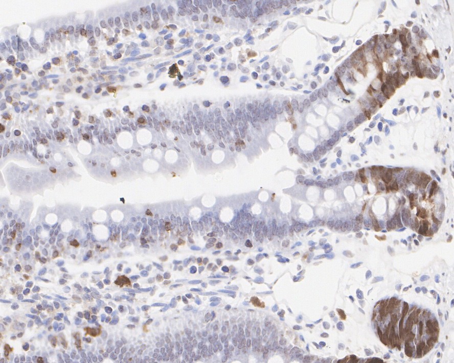
Immunohistochemical analysis of paraffin-embedded rat small intestine tissue with Rabbit anti-Phospho-Cdk2 (Y15) antibody at 1/1,000 dilution. The section was pre-treated using heat mediated antigen retrieval with sodium citrate buffer (pH 6.0) for 2 minutes. The tissues were blocked in 1% BSA for 20 minutes at room temperature, washed with ddH2O and PBS, and then probed with the primary antibody at 1/1,000 dilution for 1 hour at room temperature. The detection was performed using an HRP conjugated compact polymer system. DAB was used as the chromogen. Tissues were counterstained with hematoxylin and mounted with DPX.| Product Name | Phospho-Cdk2 (Y15) Recombinant Rabbit Monoclonal Antibody |
|---|---|
| Antibody Type | Primary Antibodies |
| Product description | In vertebrates, as in yeast, multiple cyclins have been identified, including a total of eight such regulatory proteins in mammals. In contrast to the situation in yeast, the Cdc2 p34 kinase is not the only catalytic subunit identified in vertebrates that can interact with cyclins. While Cdc2 p34 is essential for the G2 to M transition in vertebrate cells, a second Cdc2-related kinase has also been implicated in cell cycle control. This protein, designated cyclin-dependent kinase 2 (Cdk2) p33, also binds to cyclins and its kinase activity is temporally regulated during the cell cycle. Several additional Cdc2 p34-related cyclin dependent kinases have been identified. These include Cdk3-Cdk8, PCTAIRE-1-3 and KKIALRE. |
| Immunogen | Synthetic phospho-peptide corresponding to residues surrounding Tyr15 of human Cdk2. |
| Modification | p-Y15 |
| Clonality | Monoclonal |
|---|---|
| Isotype | IgG |
| Host Species | Recombinant rabbit |
| Tested Applications | IHCWB |
| WB:1:1000 IHC:1:200-1:1000 |
|
| Species Reactivity | HumanMouseRat |
| Concentration | 1mg/ml |
| Purification | Protein A |
| Alternative Names | Cdc2 related protein kinase antibody cdc2-related protein kinase antibody CDC28 antibody CDC2A antibody Cdk 2 antibody CDK1 antibody CDK2 antibody CDK2_HUMAN antibody CDKN2 antibody Cell devision kinase 2 antibody Cell division protein kinase 2 antibody Cyclin dependent kinase 2 antibody cyclin dependent kinase 2-alpha antibody Cyclin-dependent kinase 2 antibody kinase Cdc2 antibody MPF antibody p33 protein kinase antibody p33(CDK2) antibody |
|---|---|
| Molecular Weight(MW) | 34kDa |
| Cellular Localization | Cytoplasm, Cytoskeleton, Endosome, Nucleus. |
WB
Western blot analysis of Phospho-Cdk2(Y15) on Hela cell lysates. Lane 1: Hela cells, whole cell lysate, 10ug/lane, Lane 2: Hela cells treated with 2.8ug/ul lambda-PP for 30 minutes, whole cell lysates, 10ug/lane, All lanes : Anti-Phospho-Cdk2(Y15) antibody at 1/500 dilution. Anti-Cdk2 antibody at 1/500 dilution. Anti-GAPDH antibody at 1/10,000 dilution. Goat Anti-Rabbit IgG H&L (HRP)at 1/200,000 dilution. Blocking and diluting buffer: 5% BSA. Exposure time: 30 seconds.
IHC
Immunohistochemical analysis of paraffin-embedded human breast carcinoma tissue with Rabbit anti-Phospho-Cdk2 (Y15) antibody at 1/1,000 dilution. The section was pre-treated using heat mediated antigen retrieval with sodium citrate buffer (pH 6.0) for 2 minutes. The tissues were blocked in 1% BSA for 20 minutes at room temperature, washed with ddH2O and PBS, and then probed with the primary antibody at 1/1,000 dilution for 1 hour at room temperature. The detection was performed using an HRP conjugated compact polymer system. DAB was used as the chromogen. Tissues were counterstained with hematoxylin and mounted with DPX.
IHC
Immunohistochemical analysis of paraffin-embedded mouse small intestine tissue with Rabbit anti-Phospho-Cdk2 (Y15) antibody at 1/200 dilution. The section was pre-treated using heat mediated antigen retrieval with sodium citrate buffer (pH 6.0) for 2 minutes. The tissues were blocked in 1% BSA for 20 minutes at room temperature, washed with ddH2O and PBS, and then probed with the primary antibody at 1/200 dilution for 1 hour at room temperature. The detection was performed using an HRP conjugated compact polymer system. DAB was used as the chromogen. Tissues were counterstained with hematoxylin and mounted with DPX.
IHC
Immunohistochemical analysis of paraffin-embedded rat small intestine tissue with Rabbit anti-Phospho-Cdk2 (Y15) antibody at 1/1,000 dilution. The section was pre-treated using heat mediated antigen retrieval with sodium citrate buffer (pH 6.0) for 2 minutes. The tissues were blocked in 1% BSA for 20 minutes at room temperature, washed with ddH2O and PBS, and then probed with the primary antibody at 1/1,000 dilution for 1 hour at room temperature. The detection was performed using an HRP conjugated compact polymer system. DAB was used as the chromogen. Tissues were counterstained with hematoxylin and mounted with DPX.| Application Notes | WB:1:1000 IHC:1:200-1:1000 |
|---|
| Form | Liquid |
|---|---|
| Storage Instructions | Store at +4℃ after thawing. Aliquot store at -20℃ or -80℃. Avoid repeated freeze / thaw cycles. |
| Storage Buffer | 1*TBS (pH7.4), 1%BSA, 40%Glycerol. Preservative: 0.05% Sodium Azide. |


