Application
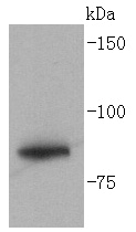
Fig1: Western blot analysis of Glycogen synthase on Hela lysates using anti- Glycogen synthase antibody at 1/1,000 dilution.Application
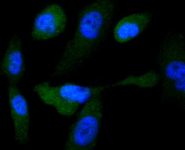
Fig2: ICC staining Glycogen synthase in PC-3M cells (green). The nuclear counter stain is DAPI (blue). Cells were fixed in paraformaldehyde, permeabilised with 0.25% Triton X100/PBS.Application
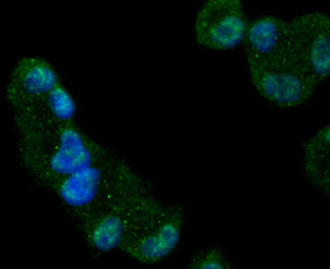
Fig3: ICC staining Glycogen synthase in Hela cells (green). The nuclear counter stain is DAPI (blue). Cells were fixed in paraformaldehyde, permeabilised with 0.25% Triton X100/PBS.Application
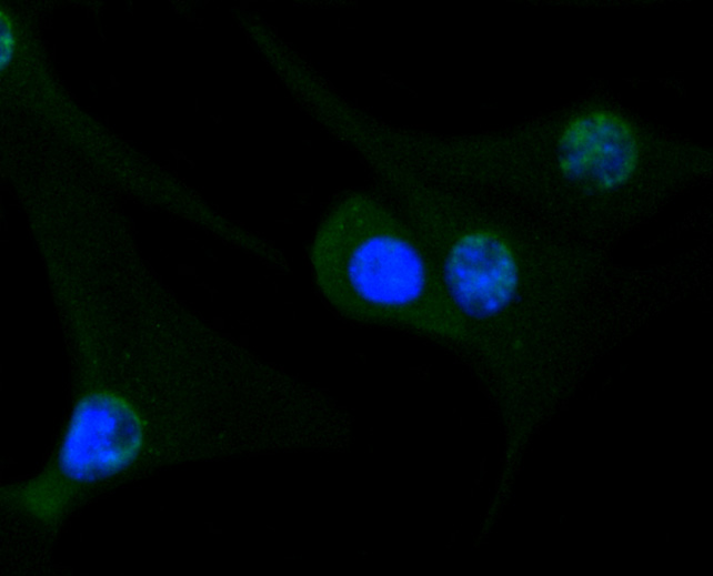
Fig4: ICC staining Glycogen synthase in NIH-3T3 cells (green). The nuclear counter stain is DAPI (blue). Cells were fixed in paraformaldehyde, permeabilised with 0.25% Triton X100/PBS.Application
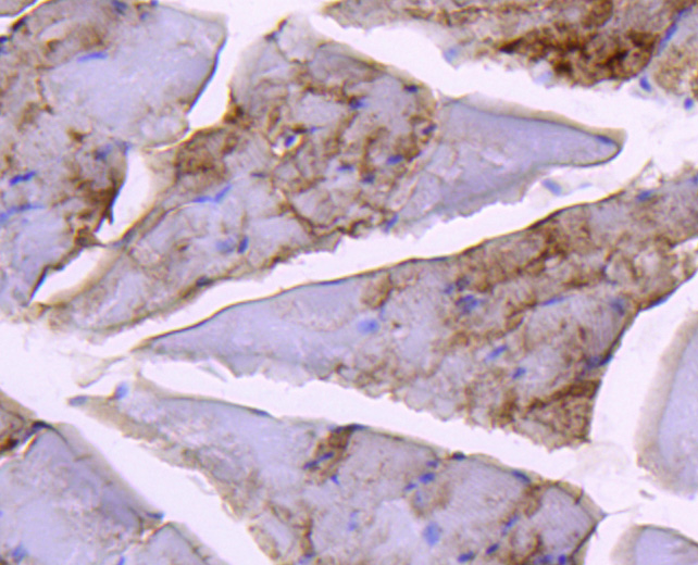
Fig5: Immunohistochemical analysis of paraffin-embedded mouse skeletal muscle tissue using anti-Glycogen synthase antibody. Counter stained with hematoxylin.Application
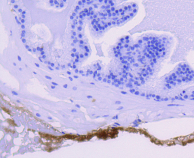
Fig6: Immunohistochemical analysis of paraffin-embedded mouse prostate tissue using anti-Glycogen synthase antibody. Counter stained with hematoxylin.Application
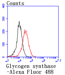
Fig7: Flow cytometric analysis of 293 cells with Glycogen synthase antibody at 1/50 dilution (red) compared with an unlabelled control (cells without incubation with primary antibody; black). Alexa Fluor 488-conjugated goat anti rabbit IgG was used as the secondary antibody.| Product Name | Glycogen synthase [SN75-05] |
|---|---|
| Antibody Type | Primary Antibodies |
| Antigen Alias | Glycogen [starch] synthase antibody Glycogen synthase 1 (muscle) antibody Glycogen synthase 1 antibody GSY antibody GYS antibody Gys1 antibody GYS1_HUMAN antibody muscle antibody |
| Product description | Glycogen [starch] synthase belongs to the mammalian/fungal glycogen synthase family of proteins. Two forms of this protein exist, a liver form and a muscle form, both of which have the same function in the glycogen biosynthesis pathway. Glycogen synthase transfers the glycosyl residue from UDP-Glucose to the nonreducing end of α-1,4-glucan. The liver glycogen synthase protein is truncated by 34 amino acids compared to the muscle form. However, these enzymes differ significantly in their amino- and carboxyl-terminal regions. Muscle glycogen synthase serves to fuel muscular activity only and is regulated by muscle contraction and by catecholamines. Liver glycogen synthase mediates blood glucose homeostasis in response to nutritional cues. Defects in the gene encoding liver glycogen synthase results in glycogen storage disease type 0 (GSD0), a rare form of fasting ketotic hypoglycemia. |
| Immunogen | recombinant protein |
| Clonality | Monoclonal |
|---|---|
| Isotype | IgG |
| Host Species | Recombinant rabbit |
| Tested Applications | WBICC/IFIHCIPFC |
| WB:1:1,000 ICC:1:50-1:200 IHC:1:50-1:200 FC:1:50-1:100 |
|
| Species Reactivity | HumanMouseRat |
| Concentration | 1mg/ml |
| Alternative Names | Glycogen [starch] synthase antibody Glycogen synthase 1 (muscle) antibody Glycogen synthase 1 antibody GSY antibody GYS antibody Gys1 antibody GYS1_HUMAN antibody muscle antibody |
|---|---|
| Molecular Weight(MW) | 84 kDa |
| Cellular Localization | Cytosol, Inclusion body, Membrane. |
Application
Fig1: Western blot analysis of Glycogen synthase on Hela lysates using anti- Glycogen synthase antibody at 1/1,000 dilution.
Application
Fig2: ICC staining Glycogen synthase in PC-3M cells (green). The nuclear counter stain is DAPI (blue). Cells were fixed in paraformaldehyde, permeabilised with 0.25% Triton X100/PBS.
Application
Fig3: ICC staining Glycogen synthase in Hela cells (green). The nuclear counter stain is DAPI (blue). Cells were fixed in paraformaldehyde, permeabilised with 0.25% Triton X100/PBS.
Application
Fig4: ICC staining Glycogen synthase in NIH-3T3 cells (green). The nuclear counter stain is DAPI (blue). Cells were fixed in paraformaldehyde, permeabilised with 0.25% Triton X100/PBS.
Application
Fig5: Immunohistochemical analysis of paraffin-embedded mouse skeletal muscle tissue using anti-Glycogen synthase antibody. Counter stained with hematoxylin.
Application
Fig6: Immunohistochemical analysis of paraffin-embedded mouse prostate tissue using anti-Glycogen synthase antibody. Counter stained with hematoxylin.
Application
Fig7: Flow cytometric analysis of 293 cells with Glycogen synthase antibody at 1/50 dilution (red) compared with an unlabelled control (cells without incubation with primary antibody; black). Alexa Fluor 488-conjugated goat anti rabbit IgG was used as the secondary antibody.| Positive Control | Hela, PC-3M, NIH-3T3, 293, mouse skeletal muscle tissue, mouse prostate tissue. |
|---|---|
| Application Notes | WB:1:1,000 ICC:1:50-1:200 IHC:1:50-1:200 FC:1:50-1:100 |
| Form | Liquid |
|---|---|
| Storage Instructions | Store at +4℃ after thawing. Aliquot store at -20℃ or -80℃. Avoid repeated freeze / thaw cycles. |
| Storage Buffer | 1*TBS (pH7.4), 1%BSA, 40%Glycerol. Preservative: 0.05% Sodium Azide. |


