Application
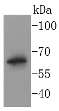
Fig1: Western blot analysis of BPI on THP-1 cells lysates using anti-BPI antibody at 1/1,000 dilution.Application
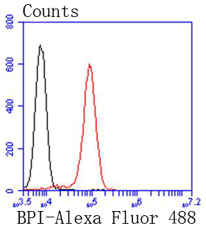
Fig2: Flow cytometric analysis of Jurkat cells with BPI antibody at 1/50 dilution (red) compared with an unlabelled control (cells without incubation with primary antibody; black). Alexa Fluor 488-conjugated goat anti rabbit IgG was used as the secondary antibody.| Product Name | BPI [SN06-63] |
|---|---|
| Antibody Type | Primary Antibodies |
| Antigen Alias | Bactericidal permeability-increasing protein antibody bactericidal/permeability-increasing protein antibody BPI antibody BPI fold containing family D, member 1 antibody BPIFD1 antibody CAP 57 antibody rBPI antibody recombinant BPI holoprotein, rBPI antibody |
| Product description | The bactericidal permeability increasing protein (BPI) is an antibacterial and endotoxin-neutralizing molecule that is abundant in the granules of polymorphonuclear leukocytes (neutrophil granules). The 31.5-kb-long human BPI gene maps to chromosome 20q11.23-q12, contains 15 exons, and encodes a 456 amino acid protein. Epithelial cells which line mucosal surfaces are the first line of defense against bacterial invasion and infection. BPI localizes to the cell surface of epithelial cells and blocks endotoxin-mediated signaling, thereby protecting mucosal surfaces against gram-negative bacteria and their endotoxin. BPI, lipopolysaccharide binding protein (LBP), phospholipid transfer protein (PLTP), and cholesteryl ester transfer protein (CETP) constitutes a family of functionally related proteins. |
| Immunogen | recombinant protein |
| Clonality | Monoclonal |
|---|---|
| Isotype | IgG |
| Host Species | Recombinant rabbit |
| Tested Applications | WBFC |
| WB:1:1,000-1:2,000 FC:1:50-1:100 |
|
| Species Reactivity | HumanZebra Fish |
| Concentration | 1mg/ml |
| Alternative Names | Bactericidal permeability-increasing protein antibody
bactericidal/permeability-increasing protein antibody
BPI antibody
BPI fold containing family D member 1 antibody BPIFD1 antibody CAP 57 antibody rBPI antibody recombinant BPI holoprotein rBPI antibody |
|---|---|
| Molecular Weight(MW) | 57 kDa |
| Cellular Localization | Secreted, Cytoplasmic granule membrane. |
| SwissProt ID | P17213 |
|---|
Application
Fig1: Western blot analysis of BPI on THP-1 cells lysates using anti-BPI antibody at 1/1,000 dilution.
Application
Fig2: Flow cytometric analysis of Jurkat cells with BPI antibody at 1/50 dilution (red) compared with an unlabelled control (cells without incubation with primary antibody; black). Alexa Fluor 488-conjugated goat anti rabbit IgG was used as the secondary antibody.| Positive Control | THP-1. |
|---|---|
| Application Notes | WB:1:1,000-1:2,000 FC:1:50-1:100 |
| Form | Liquid |
|---|---|
| Storage Instructions | Store at +4℃ after thawing. Aliquot store at -20℃ or -80℃. Avoid repeated freeze / thaw cycles. |
| Storage Buffer | 1*TBS (pH7.4), 1%BSA, 40%Glycerol. Preservative: 0.05% Sodium Azide. |


