Application
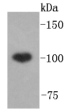
Fig1: Western blot analysis of ABCF1 on Hela lysates using anti-ABCF1 antibody at 1/1,000 dilution.Application
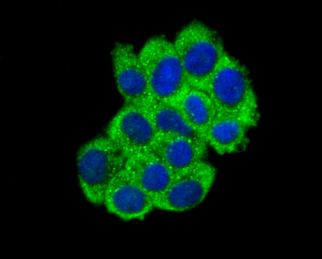
Fig2: ICC staining ABCF1 in HepG2 cells (green). The nuclear counter stain is DAPI (blue). Cells were fixed in paraformaldehyde, permeabilised with 0.25% Triton X100/PBS.Application
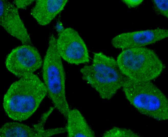
Fig3: ICC staining ABCF1 in PC-3M cells (green). The nuclear counter stain is DAPI (blue). Cells were fixed in paraformaldehyde, permeabilised with 0.25% Triton X100/PBS.Application
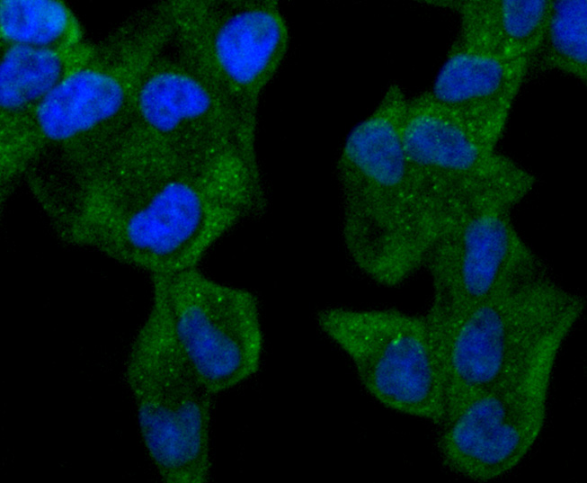
Fig4: ICC staining ABCF1 in Hela cells (green). The nuclear counter stain is DAPI (blue). Cells were fixed in paraformaldehyde, permeabilised with 0.25% Triton X100/PBS.Application
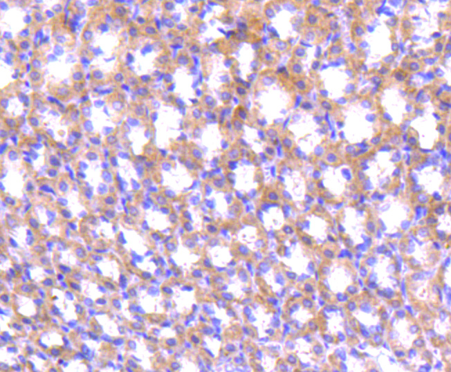
Fig5: Immunohistochemical analysis of paraffin-embedded rat stomach tissue using anti-ABCF1 antibody. Counter stained with hematoxylin.Application
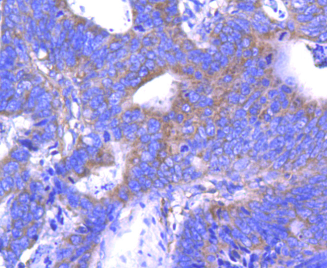
Fig6: Immunohistochemical analysis of paraffin-embedded human colon cancer tissue using anti-ABCF1 antibody. Counter stained with hematoxylin.Application
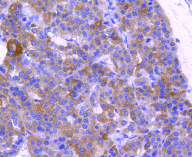
Fig7: Immunohistochemical analysis of paraffin-embedded human liver cancer tissue using anti-ABCF1 antibody. Counter stained with hematoxylin.Application
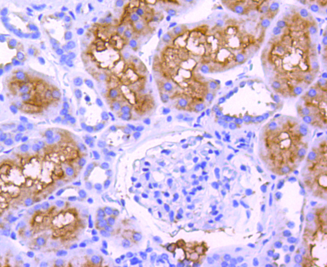
Fig8: Immunohistochemical analysis of paraffin-embedded human kidney tissue using anti-ABCF1 antibody. Counter stained with hematoxylin.| Product Name | ABCF1 [SN07-12] |
|---|---|
| Antibody Type | Primary Antibodies |
| Antigen Alias | ABC27 antibody ABC50 antibody ABCF1 antibody ABCF1_HUMAN antibody ATP binding cassette 50 (TNF alpha stimulated) antibody ATP binding cassette 50 antibody ATP binding cassette sub family F member 1 antibody ATP binding cassette, sub family F (GCN20), member 1 antibody ATP-binding cassette 50 antibody ATP-binding cassette sub-family F member 1 antibody AU041969 antibody D17Wsu166e antibody EST123147 antibody GCN20 antibody TNF alpha stimulated ABC protein antibody TNF-alpha-stimulated ABC protein antibody TNFalpha inducible ATP binding protein antibody wu:fb79c06 antibody wu:fc39a05 antibody wu:fj94a08 antibody zgc:85667 antibody |
| Product description | ABCF1 (ATP-binding cassette sub-family F member 1, TNF-alpha-stimulated ABC protein) is a 845 amino acid protein encoded by the human gene ABCF1. ABCF1 belongs to the ABC transporter family (EF3 subfamily) and contains two ABC transporter domains. ABC proteins transport various molecules across extra- and intra-cellular membranes. ABC genes are divided into seven distinct subfamilies (ABC1, MDR/TAP, MRP, ALD, OABP, GCN20, White). Unlike other members of the superfamily, this protein lacks the transmembrane domains which are characteristic of most ABC transporters. ABCF1 is believed to have a role in mRNA translation due to its interaction with eukaryotic initiation factor 2 (eIF2). It is also associated with ribosomes. ABCF1 is ubiquitously expressed and can be induced with TNF. Upon DNA damage, ABCF1 is phosphorylated by either ATM or ATR. |
| Immunogen | recombinant protein |
| Clonality | Monoclonal |
|---|---|
| Isotype | IgG |
| Host Species | Recombinant rabbit |
| Tested Applications | WBICC/IFIHC |
| WB:1:1,000-1:2,000 ICC:1:100-1:500 IHC:1:50-1:200 |
|
| Species Reactivity | HumanRat |
| Concentration | 1mg/ml |
| Alternative Names | ABC27 antibody
ABC50 antibody
ABCF1 antibody
ABCF1_HUMAN antibody
ATP binding cassette 50 (TNF alpha stimulated) antibody
ATP binding cassette 50 antibody
ATP binding cassette sub family F member 1 antibody
ATP binding cassette sub family F (GCN20) member 1 antibody ATP-binding cassette 50 antibody ATP-binding cassette sub-family F member 1 antibody AU041969 antibody D17Wsu166e antibody EST123147 antibody GCN20 antibody TNF alpha stimulated ABC protein antibody TNF-alpha-stimulated ABC protein antibody TNFalpha inducible ATP binding protein antibody wu:fb79c06 antibody wu:fc39a05 antibody wu:fj94a08 antibody zgc:85667 antibody |
|---|---|
| Molecular Weight(MW) | 96 kDa |
| Cellular Localization | Cytoplasm, Nucleus. |
Application
Fig1: Western blot analysis of ABCF1 on Hela lysates using anti-ABCF1 antibody at 1/1,000 dilution.
Application
Fig2: ICC staining ABCF1 in HepG2 cells (green). The nuclear counter stain is DAPI (blue). Cells were fixed in paraformaldehyde, permeabilised with 0.25% Triton X100/PBS.
Application
Fig3: ICC staining ABCF1 in PC-3M cells (green). The nuclear counter stain is DAPI (blue). Cells were fixed in paraformaldehyde, permeabilised with 0.25% Triton X100/PBS.
Application
Fig4: ICC staining ABCF1 in Hela cells (green). The nuclear counter stain is DAPI (blue). Cells were fixed in paraformaldehyde, permeabilised with 0.25% Triton X100/PBS.
Application
Fig5: Immunohistochemical analysis of paraffin-embedded rat stomach tissue using anti-ABCF1 antibody. Counter stained with hematoxylin.
Application
Fig6: Immunohistochemical analysis of paraffin-embedded human colon cancer tissue using anti-ABCF1 antibody. Counter stained with hematoxylin.
Application
Fig7: Immunohistochemical analysis of paraffin-embedded human liver cancer tissue using anti-ABCF1 antibody. Counter stained with hematoxylin.
Application
Fig8: Immunohistochemical analysis of paraffin-embedded human kidney tissue using anti-ABCF1 antibody. Counter stained with hematoxylin.| Positive Control | Hela, HepG2, PC-3M, rat stomach tissue, human colon cancer tissue, human liver cancer tissue, human kidney tissue. |
|---|---|
| Application Notes | WB:1:1,000-1:2,000 ICC:1:100-1:500 IHC:1:50-1:200 |
| Form | Liquid |
|---|---|
| Storage Instructions | Store at +4℃ after thawing. Aliquot store at -20℃ or -80℃. Avoid repeated freeze / thaw cycles. |
| Storage Buffer | 1*TBS (pH7.4), 1%BSA, 40%Glycerol. Preservative: 0.05% Sodium Azide. |


