Application
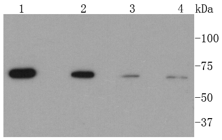
Fig1: Western blot analysis of CPT2 on different lysates using anti-CPT2 antibody at 1/1,000 dilution. Positive control: Lane 1: Hela Lane 2: 293 Lane 3: HepG2 Lane 4: NIH/3T3Application
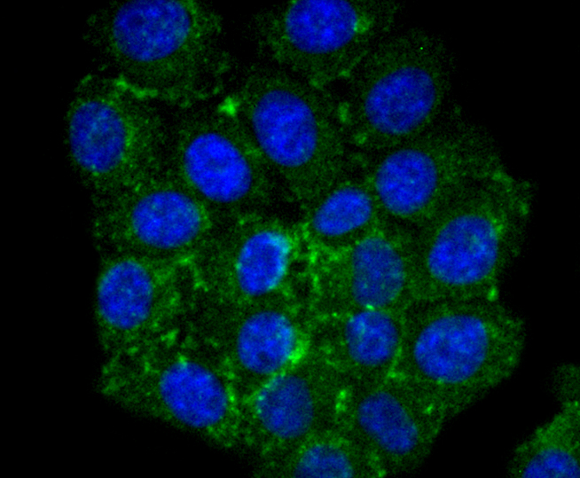
Fig2: ICC staining CPT2 in HepG2 cells (green). The nuclear counter stain is DAPI (blue). Cells were fixed in paraformaldehyde, permeabilised with 0.25% Triton X100/PBS.Application
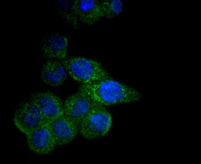
Fig3: ICC staining CPT2 in SW480 cells (green). The nuclear counter stain is DAPI (blue). Cells were fixed in paraformaldehyde, permeabilised with 0.25% Triton X100/PBS.Application
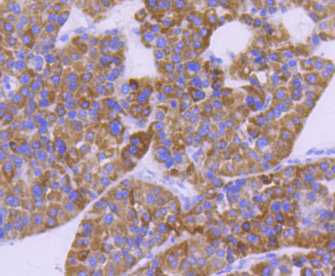
Fig4: Immunohistochemical analysis of paraffin-embedded human liver cancer tissue using anti-CPT2 antibody. Counter stained with hematoxylin.Application
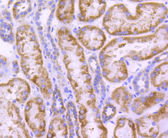
Fig5: Immunohistochemical analysis of paraffin-embedded human kidney tissue using anti-CPT2 antibody. Counter stained with hematoxylin.Application
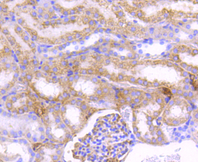
Fig6: Immunohistochemical analysis of paraffin-embedded mouse kidney tissue using anti-CPT2 antibody. Counter stained with hematoxylin.| Product Name | CPT2 [SN06-70] |
|---|---|
| Antibody Type | Primary Antibodies |
| Antigen Alias | Carnitine O palmitoyltransferase 2 antibody Carnitine O palmitoyltransferase 2 mitochondrial antibody Carnitine O-palmitoyltransferase 2 antibody Carnitine palmitoyltransferase 2 antibody Carnitine palmitoyltransferase II antibody CPT 1 antibody CPT 2 antibody CPT II antibody CPT1 antibody CPT2 antibody CPT2_HUMAN antibody CPTASE antibody CPTII antibody IIAE4 antibody mitochondrial antibody |
| Product description | The mitochondrial β-oxidation of long-chain fatty acids is initiated by the sequential action of carnitine palmitoyltransferase (CPT) I (outer membrane and detergent labile) and II (inner membrane and detergent stable), together with carnitine carrier. CPTI catalyzes the first reaction in the transport of long-chain fatty acids from the cytoplasm to the mitochondrion, a rate-limiting step in beta-oxidation. Two types of CPTI are known, the liver (CPTIA) and muscle (CPTIB) isoforms. The muscle type protein is specially expressed in heart and skeletal muscle. Membrane-bound CPTI, but not CPTII, is inhibited reversibly by malonyl-coenzyme A (CoA). Unlike CPTII, CPTI requires membrane integrity for catalytic function. In addition, glutamic acid 3 and histidine 5 are necessary for malonyl CoA inhibition and binding to liver CPTI, but not for catalytic activity. |
| Immunogen | recombinant protein |
| Clonality | Monoclonal |
|---|---|
| Isotype | IgG |
| Host Species | Recombinant rabbit |
| Tested Applications | WBICC/IFIHC |
| WB:1:500-1:1,000 ICC:1:50-1:200 IHC:1:50-1:200 |
|
| Species Reactivity | HumanMouseRat |
| Concentration | 1mg/ml |
| Alternative Names | Carnitine O palmitoyltransferase 2 antibody Carnitine O palmitoyltransferase 2 mitochondrial antibody Carnitine O-palmitoyltransferase 2 antibody Carnitine palmitoyltransferase 2 antibody Carnitine palmitoyltransferase II antibody CPT 1 antibody CPT 2 antibody CPT II antibody CPT1 antibody CPT2 antibody CPT2_HUMAN antibody CPTASE antibody CPTII antibody IIAE4 antibody mitochondrial antibody |
|---|---|
| Molecular Weight(MW) | 74 kDa |
| Cellular Localization | Mitochondrion inner membrane. |
Application
Fig1: Western blot analysis of CPT2 on different lysates using anti-CPT2 antibody at 1/1,000 dilution. Positive control: Lane 1: Hela Lane 2: 293 Lane 3: HepG2 Lane 4: NIH/3T3
Application
Fig2: ICC staining CPT2 in HepG2 cells (green). The nuclear counter stain is DAPI (blue). Cells were fixed in paraformaldehyde, permeabilised with 0.25% Triton X100/PBS.
Application
Fig3: ICC staining CPT2 in SW480 cells (green). The nuclear counter stain is DAPI (blue). Cells were fixed in paraformaldehyde, permeabilised with 0.25% Triton X100/PBS.
Application
Fig4: Immunohistochemical analysis of paraffin-embedded human liver cancer tissue using anti-CPT2 antibody. Counter stained with hematoxylin.
Application
Fig5: Immunohistochemical analysis of paraffin-embedded human kidney tissue using anti-CPT2 antibody. Counter stained with hematoxylin.
Application
Fig6: Immunohistochemical analysis of paraffin-embedded mouse kidney tissue using anti-CPT2 antibody. Counter stained with hematoxylin.| Positive Control | 293, NIH/3T3, Hela, HepG2, SW480, human liver cancer tissue, mouse kidney tissue, human kidney tissue. |
|---|---|
| Application Notes | WB:1:500-1:1,000 ICC:1:50-1:200 IHC:1:50-1:200 |
| Form | Liquid |
|---|---|
| Storage Instructions | Store at +4℃ after thawing. Aliquot store at -20℃ or -80℃. Avoid repeated freeze / thaw cycles. |
| Storage Buffer | 1*TBS (pH7.4), 1%BSA, 40%Glycerol. Preservative: 0.05% Sodium Azide. |


