WB
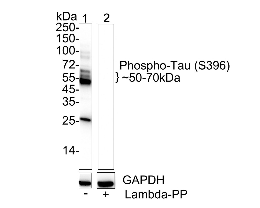
Western blot analysis of Phospho-Tau (S396) on different lysates with Rabbit anti-Phospho-Tau (S396) antibody at 1/2,000 dilution. Lane 1: Mouse brain tissue lysate, Lane 2: Mouse brain tissue lysate treated with lambda-PP for 30 minutes, Lysates/proteins at 40 µg/Lane. Exposure time: 10 seconds; 4-20% SDS-PAGE gel. Proteins were transferred to a PVDF membrane and blocked with 5% NFDM/TBST for 1 hour at room temperature. The primary antibody at 1/2,000 dilution was used in 5% NFDM/TBST at 4℃ overnight. Goat Anti-Rabbit IgG - HRP Secondary Antibody at 1/50,000 dilution was used for 1 hour at room temperature.WB
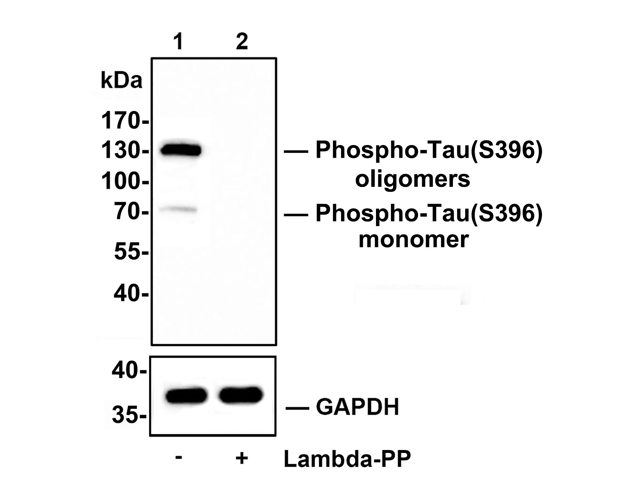
Western blot analysis of Phospho-Tau(S396) on SHSY5Y cell lysates. Lane 1: SHSY5Y cells, whole cell lysate, 10ug/lane, Lane 2: SHSY5Y cells treated with 2.8ug/ul lambda-PP for 30 minutes, whole cell lysates, 10ug/lane, All lanes : Anti-Phospho-Tau(S396) antibody at 1/500 dilution. Anti-GAPDH antibody at 1/10,000 dilution. Goat Anti-Rabbit IgG H&L (HRP) at 1/200,000 dilution. Blocking and diluting buffer: 5% BSA. Exposure time: 2 minutes 34 seconds.IHC
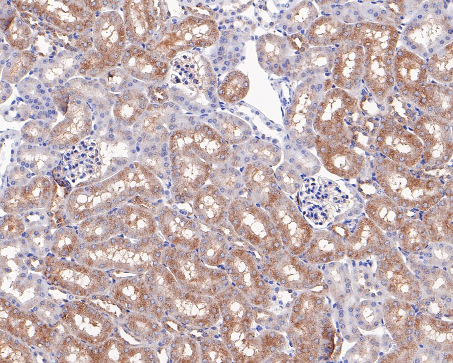
Immunohistochemical analysis of paraffin-embedded mouse kidney tissue with Rabbit anti-Phospho-Tau (S396) antibody at 1/200 dilution. The section was pre-treated using heat mediated antigen retrieval with Tris-EDTA buffer (pH 9.0) for 20 minutes. The tissues were blocked in 1% BSA for 20 minutes at room temperature, washed with ddH2O and PBS, and then probed with the primary antibody at 1/200 dilution for 1 hour at room temperature. The detection was performed using an HRP conjugated compact polymer system. DAB was used as the chromogen. Tissues were counterstained with hematoxylin and mounted with DPX.IHC
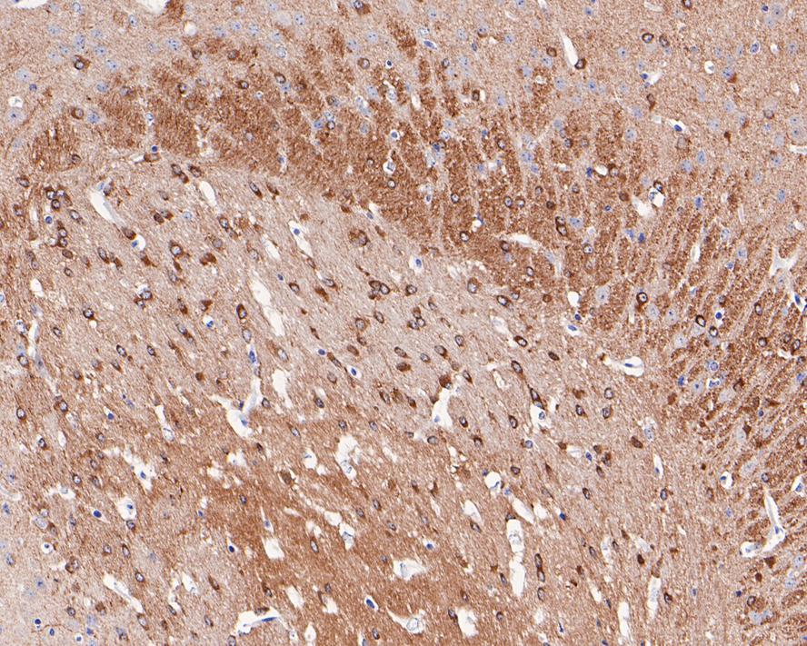
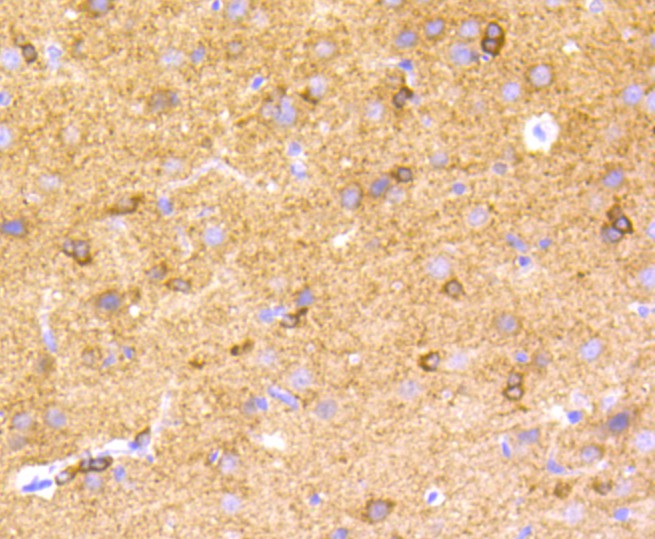
Immunohistochemical analysis of paraffin-embedded mouse brain tissue with Rabbit anti-Phospho-Tau (S396) antibody at 1/50 dilution. The section was pre-treated using heat mediated antigen retrieval with Tris-EDTA buffer (pH 9.0) for 20 minutes. The tissues were blocked in 1% BSA for 20 minutes at room temperature, washed with ddH2O and PBS, and then probed with the primary antibody at 1/50 dilution for 1 hour at room temperature. The detection was performed using an HRP conjugated compact polymer system. DAB was used as the chromogen. Tissues were counterstained with hematoxylin and mounted with DPX.IHC
Immunohistochemical analysis of paraffin-embedded rat brain tissue using anti-Phospho-Tau (S396) antibody. The section was pre-treated using heat mediated antigen retrieval with Tris-EDTA buffer (pH 8.0-8.4) for 20 minutes.The tissues were blocked in 5% BSA for 30 minutes at room temperature, washed with ddH2O and PBS, and then probed with the primary antibody (1/50) for 30 minutes at room temperature. The detection was performed using an HRP conjugated compact polymer system. DAB was used as the chromogen. Tissues were counterstained with hematoxylin and mounted with DPX.ICC/IF
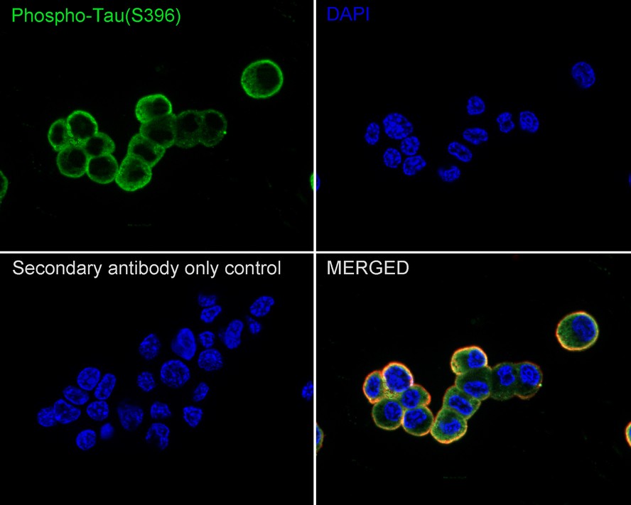
Immunocytochemistry analysis of PC-12 cells labeling Phospho-Tau (S396) with Rabbit anti-Phospho-Tau (S396) antibody at 1/100 dilution. Cells were fixed in 4% paraformaldehyde for 20 minutes at room temperature, permeabilized with 0.1% Triton X-100 in PBS for 5 minutes at room temperature, then blocked with 1% BSA in 10% negative goat serum for 1 hour at room temperature. Cells were then incubated with Rabbit anti-Phospho-Tau (S396) antibody at 1/100 dilution in 1% BSA in PBST overnight at 4 ℃. Goat Anti-Rabbit IgG H&L (488) was used as the secondary antibody at 1/1,000 dilution. PBS instead of the primary antibody was used as the secondary antibody only control. Nuclear DNA was labelled in blue with DAPI. Beta tubulin (red) was stained at 1/100 dilution overnight at +4℃. Goat Anti-Mouse IgG H&L (594) was used as the secondary antibody at 1/1,000 dilution.IF-F
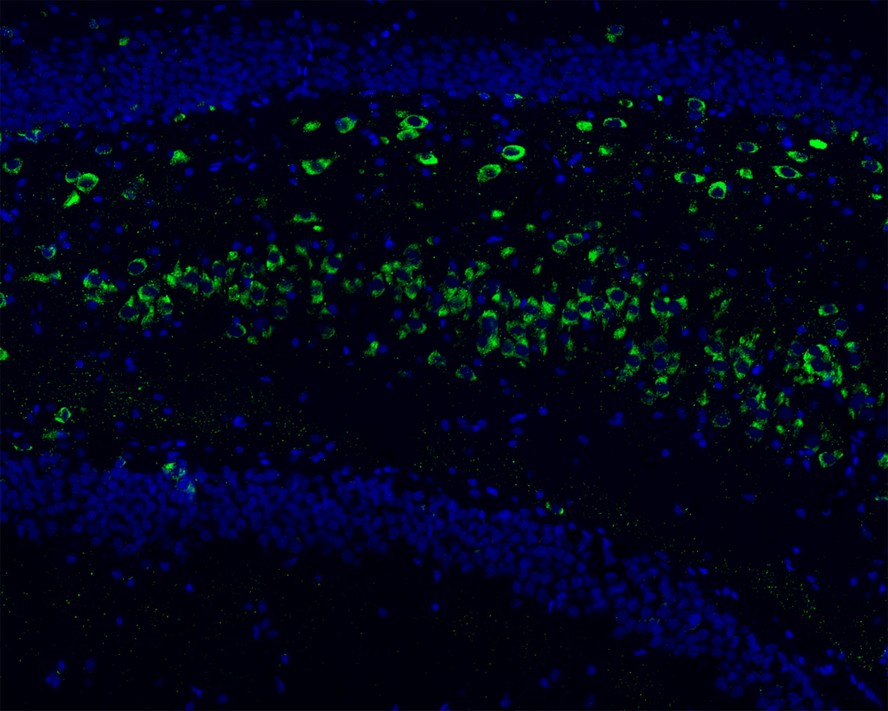
Species: Mouse, Site:Hippocampus, Sample: Frozen section, Antibody concentration: 1/200, Antigen retrieval: Not required.IP
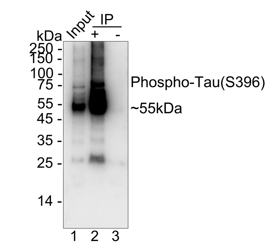
Phospho-Tau (S396) was immunoprecipitated from 0.2 mg mouse brain tissue lysate with Rabbit anti-Phospho-Tau (S396) antibody at 2 µg/10 µl beads. Western blot was performed from the immunoprecipitate using Rabbit anti-Phospho-Tau (S396) antibody at 1/1,000 dilution. HRP Conjugated Anti-Rabbit IgG for IP Nano-secondary antibody at 1/5,000 dilution was used for 1 hour at room temperature. Lane 1: Mouse brain tissue lysate (input), Lane 2: Rabbit anti-Phospho-Tau (S396) antibody IP in mouse brain tissue lysate, Lane 3: Rabbit IgG instead of Rabbit anti-Phospho-Tau (S396) antibody in mouse brain tissue ysate, Blocking/Dilution buffer: primary antibody dilution, Exposure time: 3 seconds.| Product Name | Phospho-Tau (S396) Recombinant Rabbit Monoclonal Antibody |
|---|---|
| Antibody Type | Primary Antibodies |
| Product description | Tau, also known as MAPT (microtubule-associated protein tau), MAPTL, MTBT1 or TAU, is a 758 amino acid protein that localizes to the cytoplasm, as well as to the cytoskeleton and the cell membrane, and contains four Tau/MAP repeats. Expressed in neuronal tissue and existing as multiple alternatively spliced isoforms, Tau functions to promote microtubule assembly and stability and is thought to be involved in the maintenance of neuronal polarity. Tau may also link microtubules with neural plasma membrane components and, in addition to its role in microtubule stability, is also necessary for cytoskeletal plasticity. Tau is highly subject to a variety of post-translational modifications, including phosphorylation on serine and threonine residues, polyubiquitination (and subsequent proteasomal degradation) and glycation of specific Tau isoforms. Defects in the gene encoding Tau are associated with Alzheimers disease, pallido-ponto-nigral degeneration (PPND), corticobasal degeneration (CBD) and progressive supranuclear palsy (PSP). |
| Immunogen | Synthetic phospho-peptide corresponding to residues surrounding Ser396 of human Tau. |
| Modification | p-S396 |
| Clonality | Monoclonal |
|---|---|
| Isotype | IgG |
| Host Species | Recombinant rabbit |
| Tested Applications | ICC/IFIF-FIHCIPWB |
| WB:1:500-1:2000 IHC:1:50-1:200 ICC/IF:1:100-1:500 IF-F:1:200-1:500 IP:1-2μg/sample |
|
| Species Reactivity | HumanMouseRat |
| Concentration | 1mg/ml |
| Purification | Protein A |
| Alternative Names | AI413597 antibody
AW045860 antibody
DDPAC antibody
FLJ31424 antibody
FTDP 17 antibody
G protein beta1/gamma2 subunit interacting factor 1 antibody
MAPT antibody
MAPTL antibody
MGC134287 antibody
MGC138549 antibody
MGC156663 antibody
Microtubule associated protein tau antibody
Microtubule associated protein tau isoform 4 antibody
Microtubule-associated protein tau antibody
MSTD antibody
Mtapt antibody
MTBT1 antibody
MTBT2 antibody
Neurofibrillary tangle protein antibody
Paired helical filament tau antibody
Paired helical filament-tau antibody
PHF tau antibody
PHF-tau antibody
PPND antibody
PPP1R103 antibody
Protein phosphatase 1 regulatory subunit 103 antibody pTau antibody RNPTAU antibody TAU antibody TAU_HUMAN antibody Tauopathy and respiratory failure included antibody |
|---|---|
| Molecular Weight(MW) | 79 kDa(Observed band size: 50-70,130 kDa) |
| Cellular Localization | Cytoplasm, Cell membrane, Cell projection. |
WB
Western blot analysis of Phospho-Tau (S396) on different lysates with Rabbit anti-Phospho-Tau (S396) antibody at 1/2,000 dilution. Lane 1: Mouse brain tissue lysate, Lane 2: Mouse brain tissue lysate treated with lambda-PP for 30 minutes, Lysates/proteins at 40 µg/Lane. Exposure time: 10 seconds; 4-20% SDS-PAGE gel. Proteins were transferred to a PVDF membrane and blocked with 5% NFDM/TBST for 1 hour at room temperature. The primary antibody at 1/2,000 dilution was used in 5% NFDM/TBST at 4℃ overnight. Goat Anti-Rabbit IgG - HRP Secondary Antibody at 1/50,000 dilution was used for 1 hour at room temperature.
WB
Western blot analysis of Phospho-Tau(S396) on SHSY5Y cell lysates. Lane 1: SHSY5Y cells, whole cell lysate, 10ug/lane, Lane 2: SHSY5Y cells treated with 2.8ug/ul lambda-PP for 30 minutes, whole cell lysates, 10ug/lane, All lanes : Anti-Phospho-Tau(S396) antibody at 1/500 dilution. Anti-GAPDH antibody at 1/10,000 dilution. Goat Anti-Rabbit IgG H&L (HRP) at 1/200,000 dilution. Blocking and diluting buffer: 5% BSA. Exposure time: 2 minutes 34 seconds.
IHC
Immunohistochemical analysis of paraffin-embedded mouse kidney tissue with Rabbit anti-Phospho-Tau (S396) antibody at 1/200 dilution. The section was pre-treated using heat mediated antigen retrieval with Tris-EDTA buffer (pH 9.0) for 20 minutes. The tissues were blocked in 1% BSA for 20 minutes at room temperature, washed with ddH2O and PBS, and then probed with the primary antibody at 1/200 dilution for 1 hour at room temperature. The detection was performed using an HRP conjugated compact polymer system. DAB was used as the chromogen. Tissues were counterstained with hematoxylin and mounted with DPX.
IHC
Immunohistochemical analysis of paraffin-embedded mouse brain tissue with Rabbit anti-Phospho-Tau (S396) antibody at 1/50 dilution. The section was pre-treated using heat mediated antigen retrieval with Tris-EDTA buffer (pH 9.0) for 20 minutes. The tissues were blocked in 1% BSA for 20 minutes at room temperature, washed with ddH2O and PBS, and then probed with the primary antibody at 1/50 dilution for 1 hour at room temperature. The detection was performed using an HRP conjugated compact polymer system. DAB was used as the chromogen. Tissues were counterstained with hematoxylin and mounted with DPX.
IHC
Immunohistochemical analysis of paraffin-embedded rat brain tissue using anti-Phospho-Tau (S396) antibody. The section was pre-treated using heat mediated antigen retrieval with Tris-EDTA buffer (pH 8.0-8.4) for 20 minutes.The tissues were blocked in 5% BSA for 30 minutes at room temperature, washed with ddH2O and PBS, and then probed with the primary antibody (1/50) for 30 minutes at room temperature. The detection was performed using an HRP conjugated compact polymer system. DAB was used as the chromogen. Tissues were counterstained with hematoxylin and mounted with DPX.
ICC/IF
Immunocytochemistry analysis of PC-12 cells labeling Phospho-Tau (S396) with Rabbit anti-Phospho-Tau (S396) antibody at 1/100 dilution. Cells were fixed in 4% paraformaldehyde for 20 minutes at room temperature, permeabilized with 0.1% Triton X-100 in PBS for 5 minutes at room temperature, then blocked with 1% BSA in 10% negative goat serum for 1 hour at room temperature. Cells were then incubated with Rabbit anti-Phospho-Tau (S396) antibody at 1/100 dilution in 1% BSA in PBST overnight at 4 ℃. Goat Anti-Rabbit IgG H&L (488) was used as the secondary antibody at 1/1,000 dilution. PBS instead of the primary antibody was used as the secondary antibody only control. Nuclear DNA was labelled in blue with DAPI. Beta tubulin (red) was stained at 1/100 dilution overnight at +4℃. Goat Anti-Mouse IgG H&L (594) was used as the secondary antibody at 1/1,000 dilution.
IF-F
Species: Mouse, Site:Hippocampus, Sample: Frozen section, Antibody concentration: 1/200, Antigen retrieval: Not required.
IP
Phospho-Tau (S396) was immunoprecipitated from 0.2 mg mouse brain tissue lysate with Rabbit anti-Phospho-Tau (S396) antibody at 2 µg/10 µl beads. Western blot was performed from the immunoprecipitate using Rabbit anti-Phospho-Tau (S396) antibody at 1/1,000 dilution. HRP Conjugated Anti-Rabbit IgG for IP Nano-secondary antibody at 1/5,000 dilution was used for 1 hour at room temperature. Lane 1: Mouse brain tissue lysate (input), Lane 2: Rabbit anti-Phospho-Tau (S396) antibody IP in mouse brain tissue lysate, Lane 3: Rabbit IgG instead of Rabbit anti-Phospho-Tau (S396) antibody in mouse brain tissue ysate, Blocking/Dilution buffer: primary antibody dilution, Exposure time: 3 seconds.| Application Notes | WB:1:500-1:2000 IHC:1:50-1:200 ICC/IF:1:100-1:500 IF-F:1:200-1:500 IP:1-2μg/sample |
|---|
| Form | Liquid |
|---|---|
| Storage Instructions | Store at +4℃ after thawing. Aliquot store at -20℃ or -80℃. Avoid repeated freeze / thaw cycles. |
| Storage Buffer | 1*TBS (pH7.4), 1%BSA, 40%Glycerol. Preservative: 0.05% Sodium Azide. |


