WB
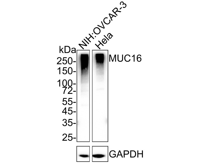
Western blot analysis of MUC16 on different lysates with Rabbit anti-MUC16 antibody at 1/1,000 dilution. Lane 1: NIH:OVCAR-3 cell lysate, Lane 2: HeLa cell lysate, Lysates/proteins at 20 µg/Lane. Exposure time: 25 seconds; 4-20% SDS-PAGE gel. Proteins were transferred to a PVDF membrane and blocked with 5% NFDM/TBST for 1 hour at room temperature. The primary antibody at 1/1,000 dilution was used in 5% NFDM/TBST at 4℃ overnight. Goat Anti-Rabbit IgG - HRP Secondary Antibody at 1/50,000 dilution was used for 1 hour at room temperature.ICC/IF
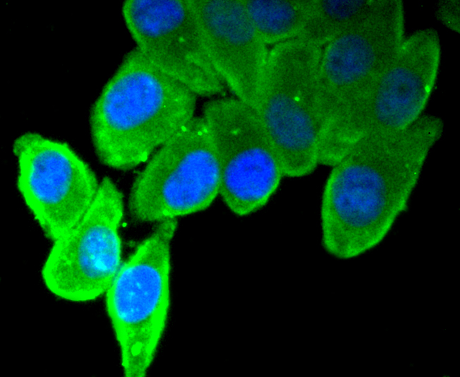
ICC staining MUC16 in HepG2 cells (green). The nuclear counter stain is DAPI (blue). Cells were fixed in paraformaldehyde, permeabilised with 0.25% Triton X100/PBS.IHC
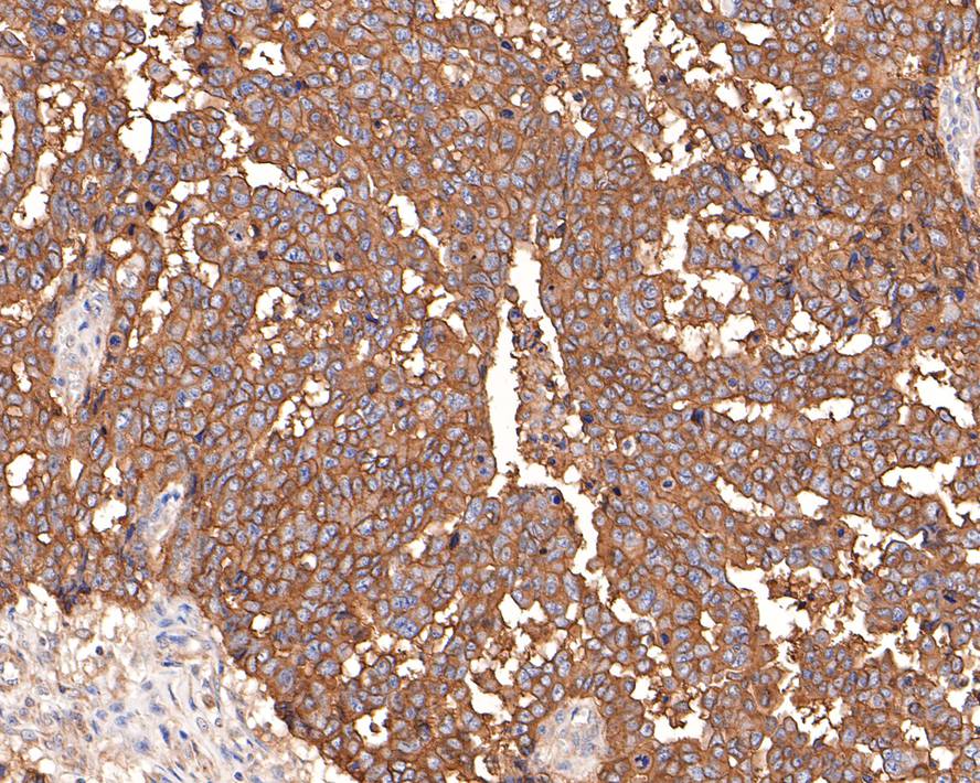
Immunohistochemical analysis of paraffin-embedded human ovarian carcinoma tissue with Rabbit anti-MUC16 antibody at 1/400 dilution. The section was pre-treated using heat mediated antigen retrieval with Tris-EDTA buffer (pH 9.0) for 20 minutes. The tissues were blocked in 1% BSA for 20 minutes at room temperature, washed with ddH2O and PBS, and then probed with the primary antibody at 1/400 dilution for 1 hour at room temperature. The detection was performed using an HRP conjugated compact polymer system. DAB was used as the chromogen. Tissues were counterstained with hematoxylin and mounted with DPX.FC
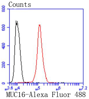
Flow cytometric analysis of Hela cells with MUC16 antibody at 1/50 dilution (red) compared with an unlabelled control (cells without incubation with primary antibody; black). Alexa Fluor 488-conjugated goat anti rabbit IgG was used as the secondary antibody| Product Name | MUC16 Recombinant Rabbit Monoclonal Antibody |
|---|---|
| Antibody Type | Primary Antibodies |
| Product description | The mucins are a family of highly glycosylated, secreted proteins with a basic structure consisting of a variable number of tandem repeats (VNTRs). Membrane-associated and secretory Mucins are high molecular weight glycoproteins of the glycocalyx (polysaccharide biofilm) that protects mucosal epithelium from particulate matter and microorganisms. Epithelial Mucins are large, secreted and cell surface glycoproteins crucial for adhesion modulation, signaling and epithelial cell protection. The number of repeats is highly polymorphic and varies among different alleles. The Mucin family consists of Mucins 1-4, Mucin 5 (AC and B), Mucins 6-8, Mucins 11-13 and Mucins 15-17. The Mucin 16 protein (also commonly referred to as CA125), encoded for by the gene MUC16, is a very high molecular weight tumor antigen consisting of three domains: a carboxy terminal domain, an extracellular domain and an amino terminal domain. Mucin 16, an ovarian cancer-associated antigen, is used as a marker to monitor the progress of epithelial ovarian cancer. It is a hydrophilic membrane-associated protein that may be involved in vitamin A functions. |
| Immunogen | Recombinant protein within Human MUC16 aa 12331-12339 / 14,507. |
| Clonality | monoclonal |
|---|---|
| Isotype | IgG |
| Host Species | Recombinant rabbit |
| Tested Applications | FCICC/IFIHCWB |
| WB:1:500-1:1,000 ICC/IF:1:100-1:500 IHC:1:100-1:500 FC:1:50-1:100 |
|
| Species Reactivity | Human |
| Concentration | 1mg/ml |
| Purification | Protein A |
| Alternative Names | CA 125 antibody CA-125 antibody CA125 antibody CA125 ovarian cancer antigen antibody Cancer antigen 125 antibody FLJ14303 antibody MUC 16 antibody MUC-16 antibody MUC16 antibody MUC16_HUMAN antibody Mucin 16 antibody Mucin 16 cell surface associated antibody Mucin-16 antibody Mucin16 antibody Ovarian cancer related tumor marker CA125 antibody Ovarian cancer-related tumor marker CA125 antibody Ovarian carcinoma antigen CA125 antibody hide |
|---|---|
| Molecular Weight(MW) | 1519kDa |
| Cellular Localization | Cell membrane, Secreted. |
| SwissProt ID | Q8WXI7 |
|---|
WB
Western blot analysis of MUC16 on different lysates with Rabbit anti-MUC16 antibody at 1/1,000 dilution. Lane 1: NIH:OVCAR-3 cell lysate, Lane 2: HeLa cell lysate, Lysates/proteins at 20 µg/Lane. Exposure time: 25 seconds; 4-20% SDS-PAGE gel. Proteins were transferred to a PVDF membrane and blocked with 5% NFDM/TBST for 1 hour at room temperature. The primary antibody at 1/1,000 dilution was used in 5% NFDM/TBST at 4℃ overnight. Goat Anti-Rabbit IgG - HRP Secondary Antibody at 1/50,000 dilution was used for 1 hour at room temperature.
ICC/IF
ICC staining MUC16 in HepG2 cells (green). The nuclear counter stain is DAPI (blue). Cells were fixed in paraformaldehyde, permeabilised with 0.25% Triton X100/PBS.
IHC
Immunohistochemical analysis of paraffin-embedded human ovarian carcinoma tissue with Rabbit anti-MUC16 antibody at 1/400 dilution. The section was pre-treated using heat mediated antigen retrieval with Tris-EDTA buffer (pH 9.0) for 20 minutes. The tissues were blocked in 1% BSA for 20 minutes at room temperature, washed with ddH2O and PBS, and then probed with the primary antibody at 1/400 dilution for 1 hour at room temperature. The detection was performed using an HRP conjugated compact polymer system. DAB was used as the chromogen. Tissues were counterstained with hematoxylin and mounted with DPX.
FC
Flow cytometric analysis of Hela cells with MUC16 antibody at 1/50 dilution (red) compared with an unlabelled control (cells without incubation with primary antibody; black). Alexa Fluor 488-conjugated goat anti rabbit IgG was used as the secondary antibody| Application Notes | WB:1:500-1:1,000 ICC/IF:1:100-1:500 IHC:1:100-1:500 FC:1:50-1:100 |
|---|
| Form | Liquid |
|---|---|
| Storage Instructions | Store at +4℃ after thawing. Aliquot store at -20℃ or -80℃. Avoid repeated freeze / thaw cycles. |
| Storage Buffer | 1*TBS (pH7.4), 1%BSA, 40%Glycerol. Preservative: 0.05% Sodium Azide. |


