WB
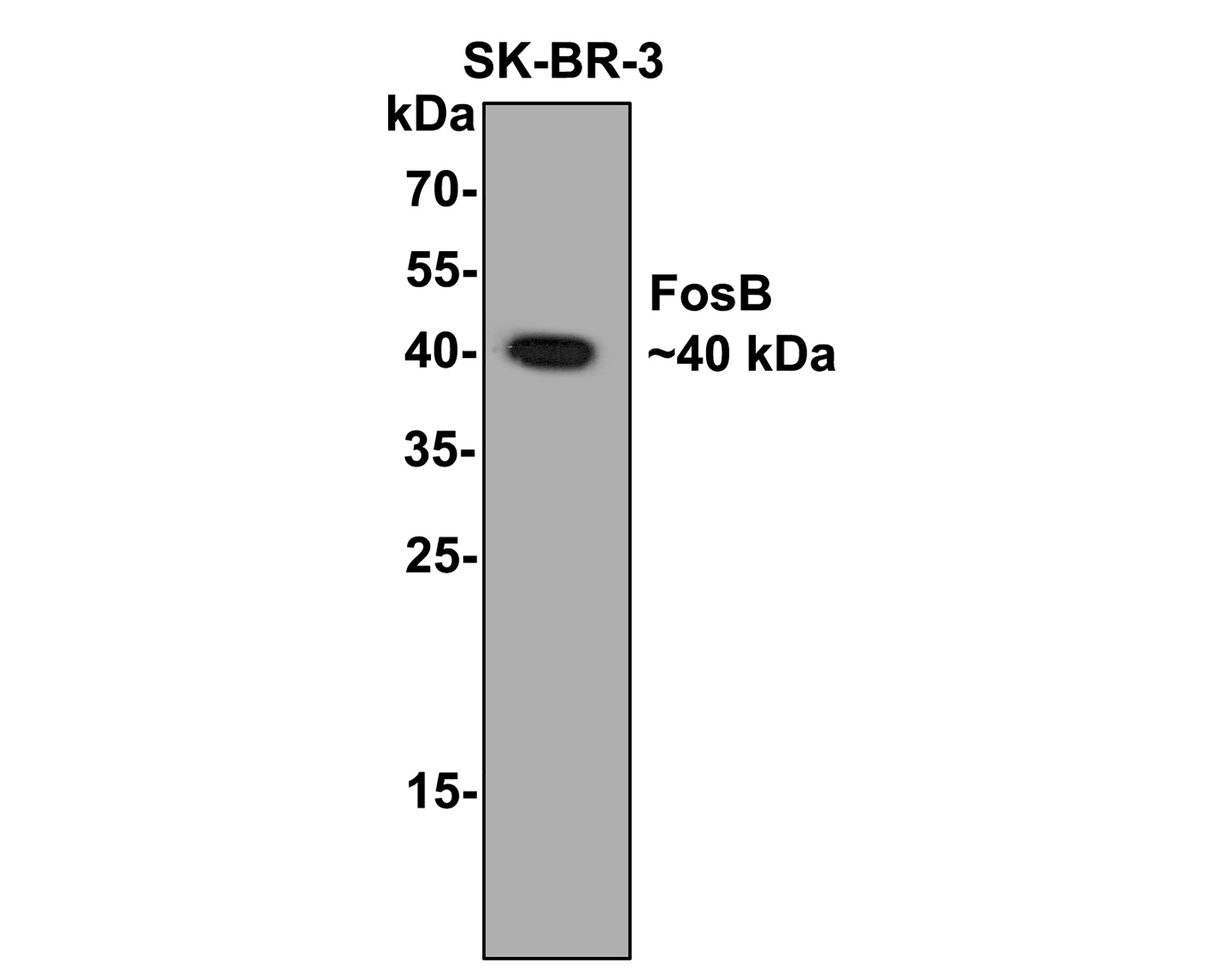
Western blot analysis of FosB on SK-Br-3 cell lysates with Rabbit anti-FosB antibody at 1/500 dilution. Lysates/proteins at 10 µg/Lane. Exposure time: 2 minutes; 12% SDS-PAGE gel. Proteins were transferred to a PVDF membrane and blocked with 5% NFDM/TBST for 1 hour at room temperature. The primary antibody at 1/500 dilution was used in 5% NFDM/TBST at room temperature for 2 hours. Goat Anti-Rabbit IgG - HRP Secondary Antibody at 1:200,000 dilution was used for 1 hour at room temperature.IHC
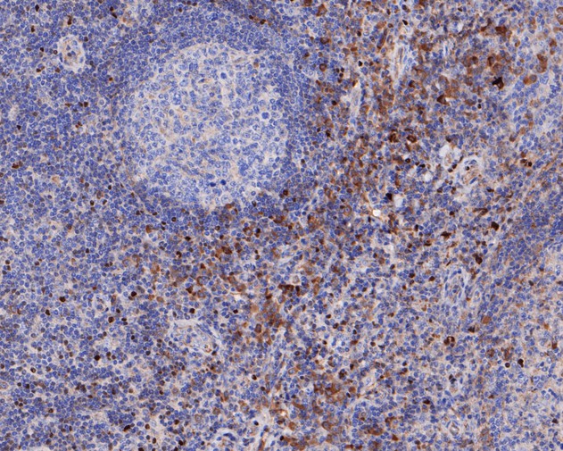
Immunohistochemical analysis of paraffin-embedded human tonsil tissue using anti-FosB antibody. The section was pre-treated using heat mediated antigen retrieval with sodium citrate buffer (pH 6.0) (high pressure) for 2 minutes. The tissues were blocked in 5% BSA for 30 minutes at room temperature, washed with ddH2O and PBS, and then probed with the primary antibody (1/50) for 30 minutes at room temperature. The detection was performed using an HRP conjugated compact polymer system. DAB was used as the chromogen. Tissues were counterstained with hematoxylin and mounted with DPX.IHC
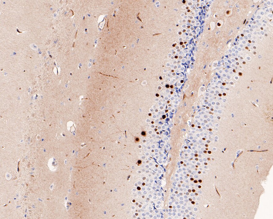
Immunohistochemical analysis of paraffin-embedded mouse hippocampus tissue with Rabbit anti-FosB antibody at 1/2,000 dilution. The section was pre-treated using heat mediated antigen retrieval with sodium citrate buffer (pH 6.0) (high pressure) for 2 minutes. The tissues were blocked in 1% BSA for 20 minutes at room temperature, washed with ddH2O and PBS, and then probed with the primary antibody at 1/2,000 dilution for 1 hour at room temperature. The detection was performed using an HRP conjugated compact polymer system. DAB was used as the chromogen. Tissues were counterstained with hematoxylin and mounted with DPX.IHC
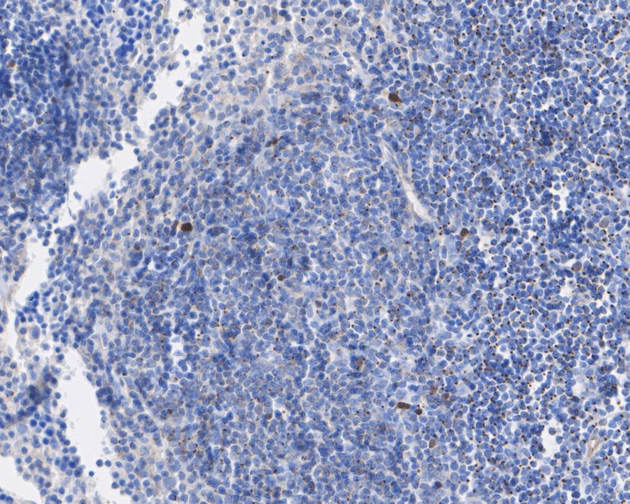
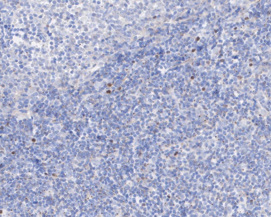
Immunohistochemical analysis of paraffin-embedded mouse spleen tissue with Rabbit anti-FosB antibody at 1/100 dilution. The section was pre-treated using heat mediated antigen retrieval with sodium citrate buffer (pH 6.0) (high pressure) for 2 minutes. The tissues were blocked in 1% BSA for 20 minutes at room temperature, washed with ddH2O and PBS, and then probed with the primary antibody at 1/100 dilution for 1 hour at room temperature. The detection was performed using an HRP conjugated compact polymer system. DAB was used as the chromogen. Tissues were counterstained with hematoxylin and mounted with DPX.IHC
Immunohistochemical analysis of paraffin-embedded rat spleen tissue with Rabbit anti-FosB antibody at 1/100 dilution. The section was pre-treated using heat mediated antigen retrieval with sodium citrate buffer (pH 6.0) (high pressure) for 2 minutes. The tissues were blocked in 1% BSA for 20 minutes at room temperature, washed with ddH2O and PBS, and then probed with the primary antibody at 1/100 dilution for 1 hour at room temperature. The detection was performed using an HRP conjugated compact polymer system. DAB was used as the chromogen. Tissues were counterstained with hematoxylin and mounted with DPX.IF-P
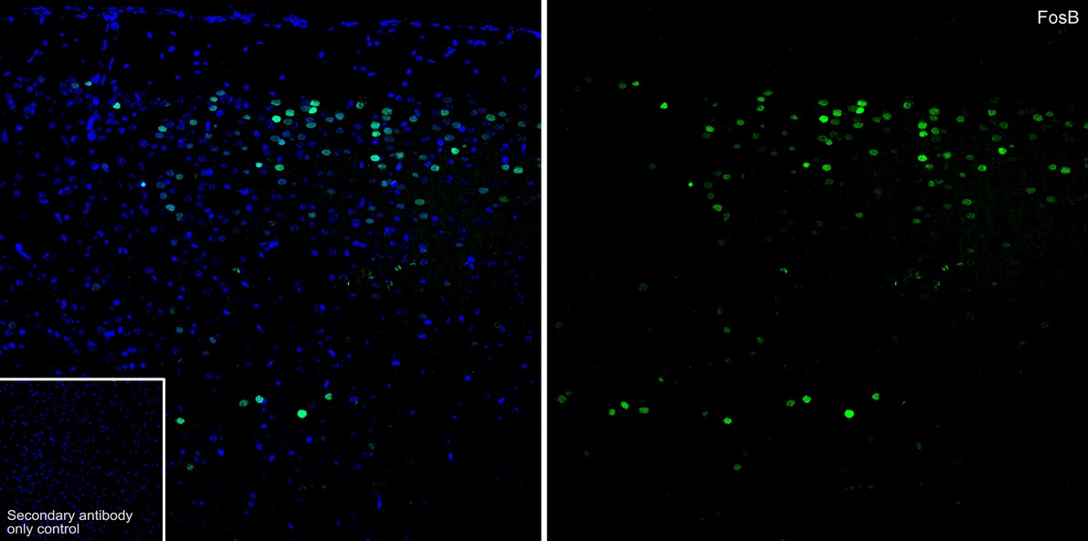
Species: Mouse, Site: brain, Sample: Paraffin-embedded section, Antibody concentration: 1/500.IF-P
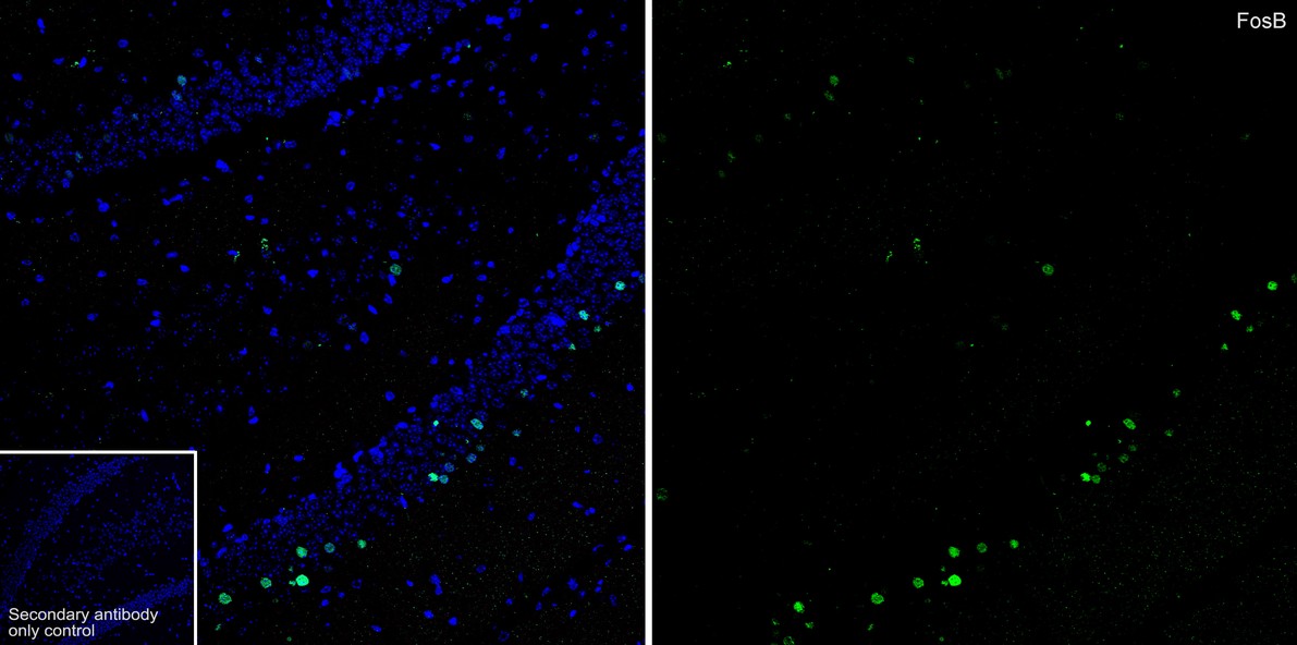
Species: Mouse, Site: hippocampus, Sample: Paraffin-embedded section, Antibody concentration: 1/500.| Product Name | FosB Recombinant Rabbit Monoclonal Antibody |
|---|---|
| Antibody Type | Primary Antibodies |
| Product description | The v-Fos oncogene was initially identified as the transforming gene of two independent murine osteosarcoma virus isolates and an avian nephroblastoma virus. The cellular homolog, c-Fos, encodes a nuclear phosphoprotein that is rapidly and transiently induced by a variety of agents and functions as a transcriptional regulator for several genes. In contrast to c-Jun proteins, which form homo- and heterodimers which bind to specific DNA TPA response elements (TREs), c-Fos proteins are only active as heterodimers with members of the Jun gene family. Murine Fos B encodes a nuclear protein of 338 amino acids which has 70% homology with c-Fos, exhibits similar kinetics of expression as c-Fos and forms heterodimers with both c-Jun and Jun B which bind to TRE DNA response elements. Functional homologs of c-Fos and Fos B include Fra-1 and Fra-2 genes. |
| Immunogen | Recombinant protein within Human FosB aa 6-189 / 338. |
| Clonality | Monoclonal |
|---|---|
| Isotype | IgG |
| Host Species | Recombinant rabbit |
| Tested Applications | IF-PIHCWB |
| WB:1:500-1:1000 IHC:1:50-1:2000 IF-P:1:500 |
|
| Species Reactivity | HumanMouseRat |
| Concentration | 1mg/ml |
| Purification | Protein A |
| Alternative Names | Activator protein 1 antibody AP 1 antibody DKFZp686C0818 antibody FBJ murine osteosarcoma viral oncogene homolog B antibody fosB antibody FOSB_HUMAN antibody G0/G1 switch regulatory protein 3 antibody G0S3 antibody GOS 3 antibody GOS B antibody GOS3 antibody GOSB antibody MGC42291 antibody Oncogene FOS B antibody Oncogene FOSB antibody Protein fos B antibody Protein fosB antibody |
|---|---|
| Molecular Weight(MW) | 36 kDa(Observed band size: 40 kDa) |
| Cellular Localization | Nucleus. |
WB
Western blot analysis of FosB on SK-Br-3 cell lysates with Rabbit anti-FosB antibody at 1/500 dilution. Lysates/proteins at 10 µg/Lane. Exposure time: 2 minutes; 12% SDS-PAGE gel. Proteins were transferred to a PVDF membrane and blocked with 5% NFDM/TBST for 1 hour at room temperature. The primary antibody at 1/500 dilution was used in 5% NFDM/TBST at room temperature for 2 hours. Goat Anti-Rabbit IgG - HRP Secondary Antibody at 1:200,000 dilution was used for 1 hour at room temperature.
IHC
Immunohistochemical analysis of paraffin-embedded human tonsil tissue using anti-FosB antibody. The section was pre-treated using heat mediated antigen retrieval with sodium citrate buffer (pH 6.0) (high pressure) for 2 minutes. The tissues were blocked in 5% BSA for 30 minutes at room temperature, washed with ddH2O and PBS, and then probed with the primary antibody (1/50) for 30 minutes at room temperature. The detection was performed using an HRP conjugated compact polymer system. DAB was used as the chromogen. Tissues were counterstained with hematoxylin and mounted with DPX.
IHC
Immunohistochemical analysis of paraffin-embedded mouse hippocampus tissue with Rabbit anti-FosB antibody at 1/2,000 dilution. The section was pre-treated using heat mediated antigen retrieval with sodium citrate buffer (pH 6.0) (high pressure) for 2 minutes. The tissues were blocked in 1% BSA for 20 minutes at room temperature, washed with ddH2O and PBS, and then probed with the primary antibody at 1/2,000 dilution for 1 hour at room temperature. The detection was performed using an HRP conjugated compact polymer system. DAB was used as the chromogen. Tissues were counterstained with hematoxylin and mounted with DPX.
IHC
Immunohistochemical analysis of paraffin-embedded mouse spleen tissue with Rabbit anti-FosB antibody at 1/100 dilution. The section was pre-treated using heat mediated antigen retrieval with sodium citrate buffer (pH 6.0) (high pressure) for 2 minutes. The tissues were blocked in 1% BSA for 20 minutes at room temperature, washed with ddH2O and PBS, and then probed with the primary antibody at 1/100 dilution for 1 hour at room temperature. The detection was performed using an HRP conjugated compact polymer system. DAB was used as the chromogen. Tissues were counterstained with hematoxylin and mounted with DPX.
IHC
Immunohistochemical analysis of paraffin-embedded rat spleen tissue with Rabbit anti-FosB antibody at 1/100 dilution. The section was pre-treated using heat mediated antigen retrieval with sodium citrate buffer (pH 6.0) (high pressure) for 2 minutes. The tissues were blocked in 1% BSA for 20 minutes at room temperature, washed with ddH2O and PBS, and then probed with the primary antibody at 1/100 dilution for 1 hour at room temperature. The detection was performed using an HRP conjugated compact polymer system. DAB was used as the chromogen. Tissues were counterstained with hematoxylin and mounted with DPX.
IF-P
Species: Mouse, Site: brain, Sample: Paraffin-embedded section, Antibody concentration: 1/500.
IF-P
Species: Mouse, Site: hippocampus, Sample: Paraffin-embedded section, Antibody concentration: 1/500.| Application Notes | WB:1:500-1:1000 IHC:1:50-1:2000 IF-P:1:500 |
|---|
| Form | Liquid |
|---|---|
| Storage Instructions | Store at +4℃ after thawing. Aliquot store at -20℃ or -80℃. Avoid repeated freeze / thaw cycles. |
| Storage Buffer | 1*TBS (pH7.4), 1%BSA, 40%Glycerol. Preservative: 0.05% Sodium Azide. |


