Application
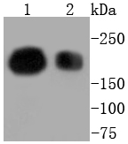
Fig1: Western blot analysis of BRG1 on different lysates using anti-BRG1 antibody at 1/1,000 dilution. Positive control: Lane 1: Jurkat Lane 2: HepG2Application
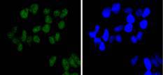
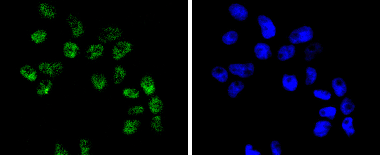
Fig2: ICC staining BRG1 in Hela cells (green). The nuclear counter stain is DAPI (blue). Cells were fixed in paraformaldehyde, permeabilised with 0.25% Triton X100/PBS.Application
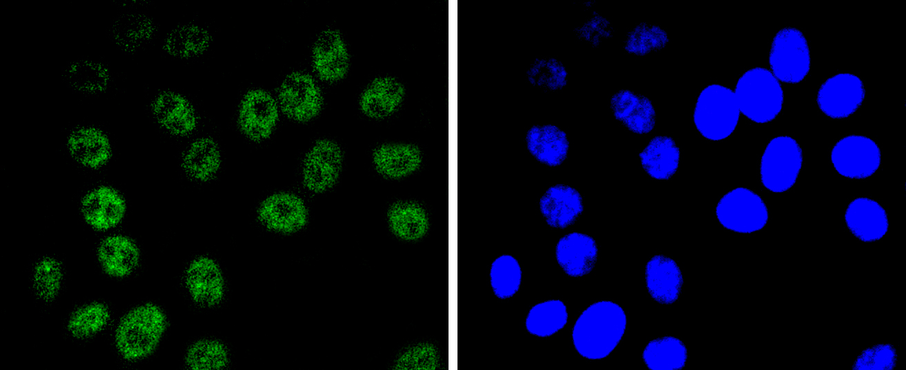
Fig3: ICC staining BRG1 in HepG2 cells (green). The nuclear counter stain is DAPI (blue). Cells were fixed in paraformaldehyde, permeabilised with 0.25% Triton X100/PBS.Application
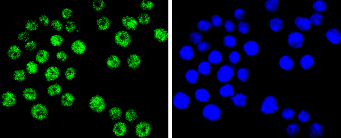
Fig4: ICC staining BRG1 in SW480 cells (green). The nuclear counter stain is DAPI (blue). Cells were fixed in paraformaldehyde, permeabilised with 0.25% Triton X100/PBS.Application
Fig5: ICC staining BRG1 in RH-35 cells (green). The nuclear counter stain is DAPI (blue). Cells were fixed in paraformaldehyde, permeabilised with 0.25% Triton X100/PBS.Application
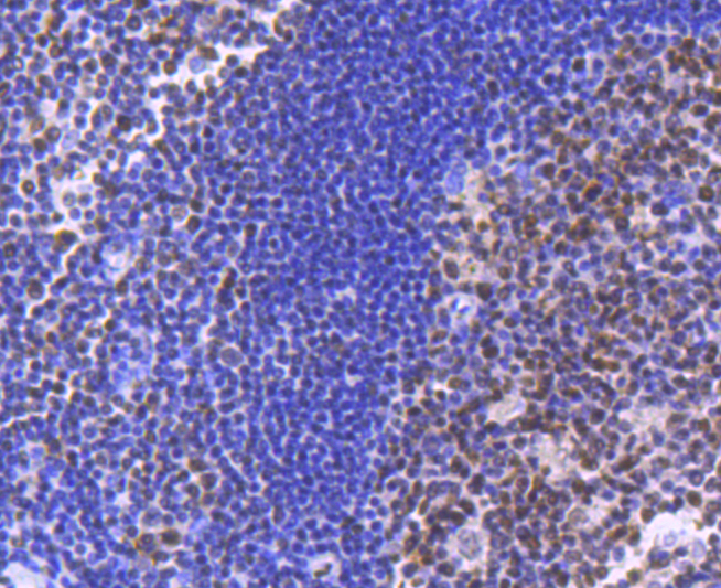
Fig6: Immunohistochemical analysis of paraffin-embedded human tonsil tissue using anti-BRG1 antibody. Counter stained with hematoxylin.Application
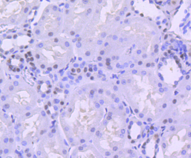
Fig7: Immunohistochemical analysis of paraffin-embedded human kidney tissue using anti-BRG1 antibody. Counter stained with hematoxylin.Application
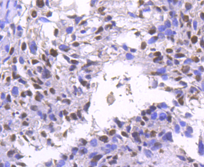
Fig8: Immunohistochemical analysis of paraffin-embedded human breast carcinoma tissue using anti-BRG1 antibody. Counter stained with hematoxylin.Application
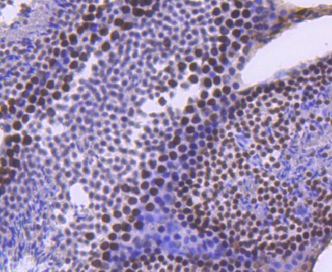
Fig9: Immunohistochemical analysis of paraffin-embedded mouse testis tissue using anti-BRG1 antibody. Counter stained with hematoxylin.Application
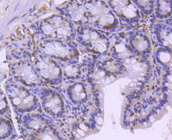
Fig10: Immunohistochemical analysis of paraffin-embedded mouse colon tissue using anti-BRG1 antibody. Counter stained with hematoxylin.Application
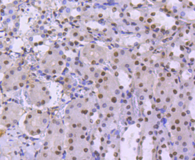
Fig11: Immunohistochemical analysis of paraffin-embedded mouse kidney tissue using anti-BRG1 antibody. Counter stained with hematoxylin.| Product Name | BRG1 [SN20-03] |
|---|---|
| Antibody Type | Primary Antibodies |
| Antigen Alias | ATP dependent helicase SMARCA4 antibody ATP-dependent helicase SMARCA4 antibody BAF 190 antibody BAF190 antibody BAF190A antibody Brahma protein like 1 antibody BRG1 antibody BRG1 associated factor 190A antibody BRG1 protein antibody BRG1-associated factor 190A antibody BRM/SWI2 related gene 1 antibody Global transcription activator homologous sequence antibody global transcription activator snf2l4 antibody Homeotic gene regulator antibody hSNF2b antibody Mitotic growth and transcription activator antibody MRD16 antibody Nuclear protein GRB1 antibody Protein brahma homolog 1 antibody Protein BRG-1 antibody Protein BRG1 antibody RTPS2 antibody SMARC A4 antibody SMARCA4 antibody SMCA4_HUMAN antibody SNF2 antibody SNF2 beta antibody SNF2 like 4 antibody SNF2-beta antibody SNF2B antibody SNF2L4 antibody SNF2LB antibody Sucrose nonfermenting like 4 antibody SWI/SNF related matrix associated actin dependent regulator of chromatin subfamily A member 4 antibody SWI/SNF related, matrix associated, actin dependent regulator of chromatin, subfamily a, member 4 antibody SWI/SNF-related matrix-associated actin-dependent regulator of chromatin subfamily A member 4 antibody SWI2 antibody Transcription activator BRG1 antibody |
| Product description | The SWI-SNF complex is involved in the activation of transcription via the remodeling of nucleosome structure in an ATP-dependent manner. Brm (also designated SNF2α) and Brg-1 (also designated SNF2β) are the ATPase subunits of the mammalian SWI-SNF complex. Brm, Brg-1, Ini1 (integrase interactor 1, also designated SNF5), BAF155 (also designated SRG3) and BAF170 are thought to comprise the functional core of the SWI-SNF complex. Addition of Ini1, BAF155 and BAF170 to Brg-1 appears to increase remodeling activity. Other complex subunits are thought to play regulatory roles. hSNF2L and hSNF2H both appear to be homologs of Drosophila ISWI, a Brm related ATPase that is present in chromatin remodeling complexes other than SWI/SNF, including the NURF (nucleosome remodeling factor). |
| Immunogen | recombinant protein |
| Clonality | Monoclonal |
|---|---|
| Isotype | IgG |
| Host Species | Recombinant rabbit |
| Tested Applications | WBICC/IFIHCIP |
| WB:1:1,000-1:5,000 ICC:1:50-1:200 IHC:1:50-1:200 |
|
| Species Reactivity | HumanMouseRat |
| Concentration | 1mg/ml |
| Alternative Names | ATP dependent helicase SMARCA4 antibody
ATP-dependent helicase SMARCA4 antibody
BAF 190 antibody
BAF190 antibody
BAF190A antibody
Brahma protein like 1 antibody
BRG1 antibody
BRG1 associated factor 190A antibody
BRG1 protein antibody
BRG1-associated factor 190A antibody
BRM/SWI2 related gene 1 antibody
Global transcription activator homologous sequence antibody
global transcription activator snf2l4 antibody
Homeotic gene regulator antibody
hSNF2b antibody
Mitotic growth and transcription activator antibody
MRD16 antibody
Nuclear protein GRB1 antibody
Protein brahma homolog 1 antibody
Protein BRG-1 antibody
Protein BRG1 antibody
RTPS2 antibody
SMARC A4 antibody
SMARCA4 antibody
SMCA4_HUMAN antibody
SNF2 antibody
SNF2 beta antibody
SNF2 like 4 antibody
SNF2-beta antibody
SNF2B antibody
SNF2L4 antibody
SNF2LB antibody
Sucrose nonfermenting like 4 antibody
SWI/SNF related matrix associated actin dependent regulator of chromatin subfamily A member 4 antibody
SWI/SNF related matrix associated actin dependent regulator of chromatin subfamily a member 4 antibody SWI/SNF-related matrix-associated actin-dependent regulator of chromatin subfamily A member 4 antibody SWI2 antibody Transcription activator BRG1 antibody |
|---|---|
| Molecular Weight(MW) | 185 kDa |
| Cellular Localization | Nucleus. |
Application
Fig1: Western blot analysis of BRG1 on different lysates using anti-BRG1 antibody at 1/1,000 dilution. Positive control: Lane 1: Jurkat Lane 2: HepG2
Application
Fig2: ICC staining BRG1 in Hela cells (green). The nuclear counter stain is DAPI (blue). Cells were fixed in paraformaldehyde, permeabilised with 0.25% Triton X100/PBS.
Application
Fig3: ICC staining BRG1 in HepG2 cells (green). The nuclear counter stain is DAPI (blue). Cells were fixed in paraformaldehyde, permeabilised with 0.25% Triton X100/PBS.
Application
Fig4: ICC staining BRG1 in SW480 cells (green). The nuclear counter stain is DAPI (blue). Cells were fixed in paraformaldehyde, permeabilised with 0.25% Triton X100/PBS.
Application
Fig5: ICC staining BRG1 in RH-35 cells (green). The nuclear counter stain is DAPI (blue). Cells were fixed in paraformaldehyde, permeabilised with 0.25% Triton X100/PBS.
Application
Fig6: Immunohistochemical analysis of paraffin-embedded human tonsil tissue using anti-BRG1 antibody. Counter stained with hematoxylin.
Application
Fig7: Immunohistochemical analysis of paraffin-embedded human kidney tissue using anti-BRG1 antibody. Counter stained with hematoxylin.
Application
Fig8: Immunohistochemical analysis of paraffin-embedded human breast carcinoma tissue using anti-BRG1 antibody. Counter stained with hematoxylin.
Application
Fig9: Immunohistochemical analysis of paraffin-embedded mouse testis tissue using anti-BRG1 antibody. Counter stained with hematoxylin.
Application
Fig10: Immunohistochemical analysis of paraffin-embedded mouse colon tissue using anti-BRG1 antibody. Counter stained with hematoxylin.
Application
Fig11: Immunohistochemical analysis of paraffin-embedded mouse kidney tissue using anti-BRG1 antibody. Counter stained with hematoxylin.| Positive Control | Jurkat, SW480, Hela, HepG2, RH-35, human breast carcinoma tissue, mouse testis tissue, human tonsil tissue, mouse colon tissue, human kidney tissue, mouse kidney tissue. |
|---|---|
| Application Notes | WB:1:1,000-1:5,000 ICC:1:50-1:200 IHC:1:50-1:200 |
| Form | Liquid |
|---|---|
| Storage Instructions | Store at +4℃ after thawing. Aliquot store at -20℃ or -80℃. Avoid repeated freeze / thaw cycles. |
| Storage Buffer | 1*TBS (pH7.4), 1%BSA, 40%Glycerol. Preservative: 0.05% Sodium Azide. |


