Application
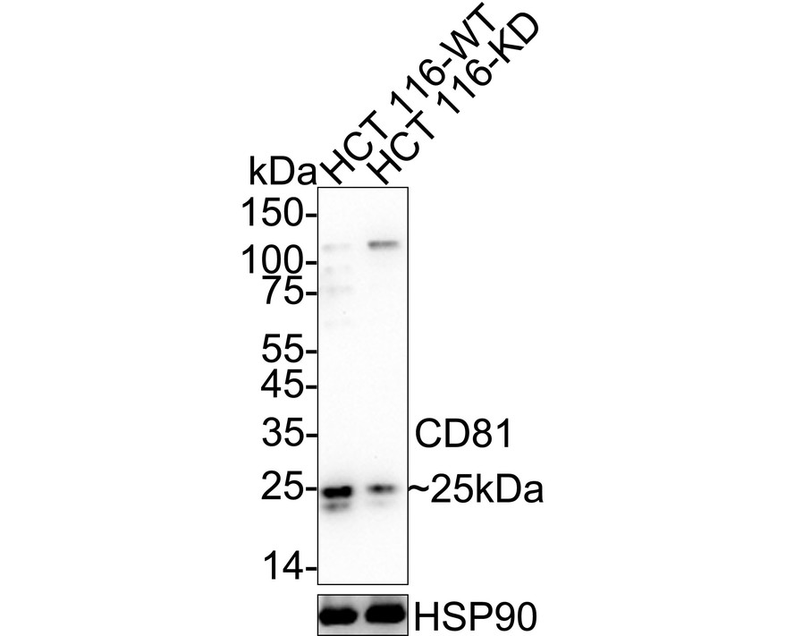
Fig1: Western blot analysis of TAPA1/CD81 on different lysates with Rabbit anti-TAPA1/CD81 antibody at 1/1,000 dilution.Application
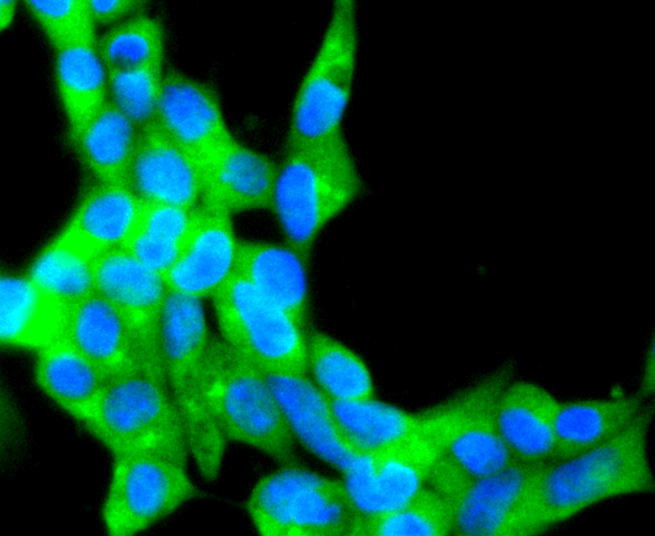
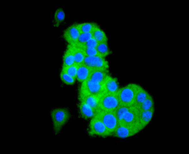
Fig2: ICC staining CD81 in 293 cells (green). The nuclear counter stain is DAPI (blue). Cells were fixed in paraformaldehyde, permeabilised with 0.25% Triton X100/PBS.Application
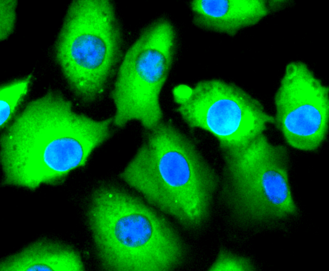
Fig3: ICC staining CD81 in A549 cells (green). The nuclear counter stain is DAPI (blue). Cells were fixed in paraformaldehyde, permeabilised with 0.25% Triton X100/PBS.Application
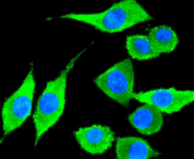
Fig4: ICC staining CD81 in SH-SY-5Y cells (green). The nuclear counter stain is DAPI (blue). Cells were fixed in paraformaldehyde, permeabilised with 0.25% Triton X100/PBS.Application
Fig5: ICC staining CD81 in PC-12 cells (green). The nuclear counter stain is DAPI (blue). Cells were fixed in paraformaldehyde, permeabilised with 0.25% Triton X100/PBS.Application
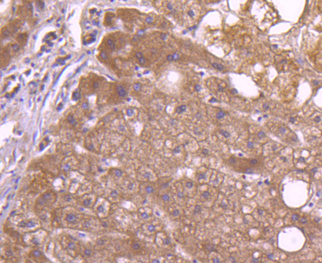
Fig6: Immunohistochemical analysis of paraffin-embedded human liver tissue using anti-CD81 antibody. Counter stained with hematoxylin.Application
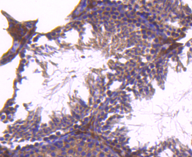
Fig7: Immunohistochemical analysis of paraffin-embedded mouse testis tissue using anti-CD81 antibody. Counter stained with hematoxylin.Application
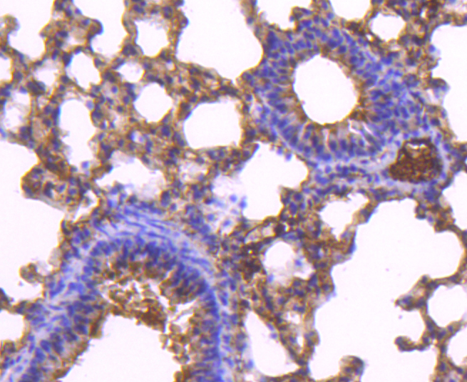
Fig8: Immunohistochemical analysis of paraffin-embedded mouse lung tissue using anti-CD81 antibody. Counter stained with hematoxylin.Application
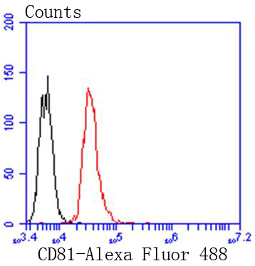
Fig9: Flow cytometric analysis of Jurkat cells with CD81 antibody at 1/50 dilution (red) compared with an unlabelled control (cells without incubation with primary antibody; black). Alexa Fluor 488-conjugated goat anti rabbit IgG was used as the secondary antibody.| Product Name | TAPA1/CD81 Recombinant Rabbit Monoclonal Antibody |
|---|---|
| Antibody Type | Primary Antibodies |
| Product description | CD81, also called TAPA-1, is a type III transmembrane protein that is broadly expressed on cells of hematopoietic, neuroectodermal and mesenchymal origin. CD81 is believed to be involved in both cell growth and signal transduction. It can be present as a multimolecular complex in association with CD37 and/or CD53, or on the surface of B cells in association with CD19, CD21 and/or MHC class II antigens. |
| Immunogen | recombinant protein |
| Isotype | IgG |
|---|---|
| Host Species | Recombinant rabbit |
| Tested Applications | FCICCIHCWB |
| WB:1:1,000-1:2,000 ICC:1:100-1:500 IHC:1:50-1:200 FC:1:50-1:100 |
|
| Species Reactivity | HumanMouseRat |
| Concentration | 1mg/ml |
| Purification | Protein A |
| Alternative Names | 26 kDa cell surface protein TAPA 1 antibody 26 kDa cell surface protein TAPA-1 antibody 26 kDa cell surface protein TAPA1 antibody CD 81 antibody CD81 antibody CD81 antigen (target of antiproliferative antibody 1) antibody CD81 antigen antibody CD81 molecule antibody CD81_HUMAN antibody CVID6 antibody S5.7 antibody TAPA 1 antibody TAPA1 antibody Target of the antiproliferative antibody 1 antibody Tetraspanin 28 antibody Tetraspanin-28 antibody Tetraspanin28 antibody Tspan 28 antibody Tspan-28 antibody Tspan28 antibody |
|---|---|
| Molecular Weight(MW) | 25 kDa |
| Cellular Localization | Membrane. |
Application
Fig1: Western blot analysis of TAPA1/CD81 on different lysates with Rabbit anti-TAPA1/CD81 antibody at 1/1,000 dilution.
Application
Fig2: ICC staining CD81 in 293 cells (green). The nuclear counter stain is DAPI (blue). Cells were fixed in paraformaldehyde, permeabilised with 0.25% Triton X100/PBS.
Application
Fig3: ICC staining CD81 in A549 cells (green). The nuclear counter stain is DAPI (blue). Cells were fixed in paraformaldehyde, permeabilised with 0.25% Triton X100/PBS.
Application
Fig4: ICC staining CD81 in SH-SY-5Y cells (green). The nuclear counter stain is DAPI (blue). Cells were fixed in paraformaldehyde, permeabilised with 0.25% Triton X100/PBS.
Application
Fig5: ICC staining CD81 in PC-12 cells (green). The nuclear counter stain is DAPI (blue). Cells were fixed in paraformaldehyde, permeabilised with 0.25% Triton X100/PBS.
Application
Fig6: Immunohistochemical analysis of paraffin-embedded human liver tissue using anti-CD81 antibody. Counter stained with hematoxylin.
Application
Fig7: Immunohistochemical analysis of paraffin-embedded mouse testis tissue using anti-CD81 antibody. Counter stained with hematoxylin.
Application
Fig8: Immunohistochemical analysis of paraffin-embedded mouse lung tissue using anti-CD81 antibody. Counter stained with hematoxylin.
Application
Fig9: Flow cytometric analysis of Jurkat cells with CD81 antibody at 1/50 dilution (red) compared with an unlabelled control (cells without incubation with primary antibody; black). Alexa Fluor 488-conjugated goat anti rabbit IgG was used as the secondary antibody.| Positive Control | JAR, PC-12, K562, A549, SH-SY-5Y, mouse lung tissue, human liver tissue, mouse testis tissue. |
|---|---|
| Application Notes | WB:1:1,000-1:2,000 ICC:1:100-1:500 IHC:1:50-1:200 FC:1:50-1:100 |
| Form | Liquid |
|---|---|
| Storage Instructions | Store at +4℃ after thawing. Aliquot store at -20℃ or -80℃. Avoid repeated freeze / thaw cycles. |
| Storage Buffer | 1*TBS (pH7.4), 1%BSA, 40%Glycerol. Preservative: 0.05% Sodium Azide. |


