Application
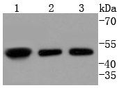
Fig1: Western blot analysis of E2F2 on different lysates using anti-E2F2 antibody at 1/1,000 dilution. Positive control: Lane 1: K562 Lane 2: 293T Lane 3: HepG2Application
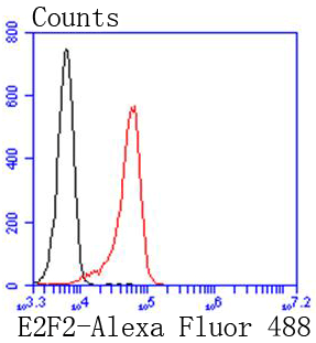
Fig2: Flow cytometric analysis of K562 cells with E2F2 antibody at 1/50 dilution (red) compared with an unlabelled control (cells without incubation with primary antibody; black). Alexa Fluor 488-conjugated goat anti rabbit IgG was used as the secondary antibody| Product Name | E2F2 [SN201-04] |
|---|---|
| Antibody Type | Primary Antibodies |
| Antigen Alias | dE2F2 antibody E2F transcription factor 2 antibody E2F-2 antibody E2F2 antibody E2F2_HUMAN antibody Transcription factor E2F2 antibody |
| Product description | The human retinoblastoma gene product appears to play an important role in the negative regulation of cell proliferation. Functional inactivation of Rb can be mediated either through mutation or as a consequence of interaction with DNA tumor virus-encoded proteins. Of all the Rb associations described to date, the identification of a complex between Rb and the transcription factor E2F most directly implicates Rb in regulation of cell proliferation. E2F was originally identified through its role in transcriptional activation of the adenovirus E2 promoter. Sequences homologous to the E2F binding site have been found upstream of a number of genes that encode proteins with putative functions in the G1 and S phases of the cell cycle. E2F-1 is a member of a broader family of transcription regulators including E2F-2, E2F-3, E2F-4, E2F-5, E2F-6 and E2F-7 each of which forms heterodimers with a second protein, DP-1, forming an “active” E2F transcriptional regulatory complex. |
| Immunogen | recombinant protein |
| Clonality | Monoclonal |
|---|---|
| Isotype | IgG |
| Host Species | Recombinant rabbit |
| Tested Applications | WBFC |
| WB:1:1,000-1:2,000 FC:1:50-1:100 |
|
| Species Reactivity | HumanMouseRat |
| Concentration | 1mg/ml |
| Alternative Names | dE2F2 antibody E2F transcription factor 2 antibody E2F-2 antibody E2F2 antibody E2F2_HUMAN antibody Transcription factor E2F2 antibody |
|---|---|
| Molecular Weight(MW) | 48 kDa |
| Cellular Localization | Nucleus. |
Application
Fig1: Western blot analysis of E2F2 on different lysates using anti-E2F2 antibody at 1/1,000 dilution. Positive control: Lane 1: K562 Lane 2: 293T Lane 3: HepG2
Application
Fig2: Flow cytometric analysis of K562 cells with E2F2 antibody at 1/50 dilution (red) compared with an unlabelled control (cells without incubation with primary antibody; black). Alexa Fluor 488-conjugated goat anti rabbit IgG was used as the secondary antibody| Positive Control | K562, 293T, HepG2. |
|---|---|
| Application Notes | WB:1:1,000-1:2,000 FC:1:50-1:100 |
| Form | Liquid |
|---|---|
| Storage Instructions | Store at +4℃ after thawing. Aliquot store at -20℃ or -80℃. Avoid repeated freeze / thaw cycles. |
| Storage Buffer | 1*TBS (pH7.4), 1%BSA, 40%Glycerol. Preservative: 0.05% Sodium Azide. |


