Application
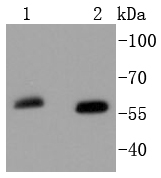
Fig1: Western blot analysis of SOX10 on different lysates using anti-SOX10 antibody at 1/1,000 dilution. Positive control: Lane 1: Mouse brain Lane 2: HepG2Application
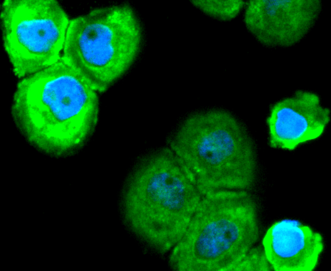
Fig2: ICC staining SOX10 in A431 cells (green). The nuclear counter stain is DAPI (blue). Cells were fixed in paraformaldehyde, permeabilised with 0.25% Triton X100/PBS.Application
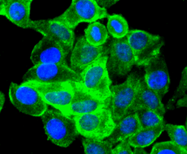
Fig3: ICC staining SOX10 in HepG2 cells (green). The nuclear counter stain is DAPI (blue). Cells were fixed in paraformaldehyde, permeabilised with 0.25% Triton X100/PBS.Application
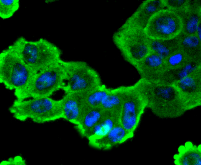
Fig4: ICC staining SOX10 in RH-35 cells (green). The nuclear counter stain is DAPI (blue). Cells were fixed in paraformaldehyde, permeabilised with 0.25% Triton X100/PBS.| Product Name | SOX10 [SN20-41] |
|---|---|
| Antibody Type | Primary Antibodies |
| Antigen Alias | DOM antibody DOM antibody Dominant megacolon mouse human homolog of antibody MGC15649 antibody PCWH antibody SOX 10 antibody SOX10 antibody SOX10_HUMAN antibody SRY (sex determining region Y) box 10 antibody SRY (sex determining region Y) box 10 antibody SRY box 10 antibody SRY box containing gene 10 antibody SRY related HMG box gene 10 antibody SRY related HMG box gene 10 antibody Transcription factor SOX 10 antibody Transcription factor SOX-10 antibody WS2E antibody WS4 antibody WS4C antibody |
| Product description | Sox genes comprise a family of genes that are related to the mammalian sex determining gene SRY. These genes similarly contain sequences that encode for the HMG-box domain, which is responsible for the sequence-specific DNA-binding activity. Sox genes encode putative transcriptional regulators implicated in the decision of cell fates during development and the control of diverse developmental processes. The highly complex group of Sox genes cluster at least 40 different loci that rapidly diverged in various animal lineages. At present, 30 Sox genes have been identified. Members of this family have been shown to be conserved during evolution and to play key roles during animal development. Some are involved in human diseases, including sex reversal. |
| Immunogen | recombinant protein |
| Clonality | Monoclonal |
|---|---|
| Isotype | IgG |
| Host Species | Recombinant rabbit |
| Tested Applications | WBICC/IF |
| WB:1:1,000-1:2,000 ICC:1:100-1:500 |
|
| Species Reactivity | HumanMouseRat |
| Concentration | 1mg/ml |
| Alternative Names | DOM antibody DOM antibody Dominant megacolon mouse human homolog of antibody MGC15649 antibody PCWH antibody SOX 10 antibody SOX10 antibody SOX10_HUMAN antibody SRY (sex determining region Y) box 10 antibody SRY (sex determining region Y) box 10 antibody SRY box 10 antibody SRY box containing gene 10 antibody SRY related HMG box gene 10 antibody SRY related HMG box gene 10 antibody Transcription factor SOX 10 antibody Transcription factor SOX-10 antibody WS2E antibody WS4 antibody WS4C antibody |
|---|---|
| Molecular Weight(MW) | 60 kDa |
| Cellular Localization | Cytoplasm, Nucleus. |
Application
Fig1: Western blot analysis of SOX10 on different lysates using anti-SOX10 antibody at 1/1,000 dilution. Positive control: Lane 1: Mouse brain Lane 2: HepG2
Application
Fig2: ICC staining SOX10 in A431 cells (green). The nuclear counter stain is DAPI (blue). Cells were fixed in paraformaldehyde, permeabilised with 0.25% Triton X100/PBS.
Application
Fig3: ICC staining SOX10 in HepG2 cells (green). The nuclear counter stain is DAPI (blue). Cells were fixed in paraformaldehyde, permeabilised with 0.25% Triton X100/PBS.
Application
Fig4: ICC staining SOX10 in RH-35 cells (green). The nuclear counter stain is DAPI (blue). Cells were fixed in paraformaldehyde, permeabilised with 0.25% Triton X100/PBS.| Positive Control | A431, MCF-7, RH-35, HepG2, mouse brain tissue. |
|---|---|
| Application Notes | WB:1:1,000-1:2,000 ICC:1:100-1:500 |
| Form | Liquid |
|---|---|
| Storage Instructions | Store at +4℃ after thawing. Aliquot store at -20℃ or -80℃. Avoid repeated freeze / thaw cycles. |
| Storage Buffer | 1*TBS (pH7.4), 1%BSA, 40%Glycerol. Preservative: 0.05% Sodium Azide. |


