Application
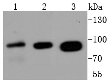
Fig1: Western blot analysis of Insulin Receptor on different lysates using anti-Insulin Receptor antibody at 1/1,000 dilution. Positive control: Lane 1: 293 Lane 2: MCF-7 Lane 3: HelaApplication
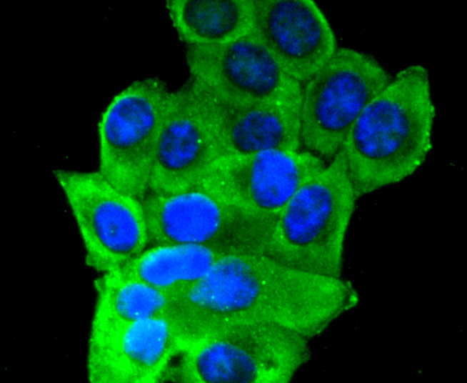
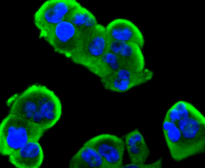
Fig2: ICC staining Insulin Receptor in HepG2 cells (green). The nuclear counter stain is DAPI (blue). Cells were fixed in paraformaldehyde, permeabilised with 0.25% Triton X100/PBS.Application
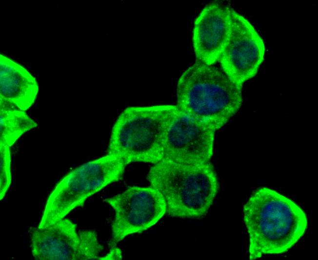
Fig3: ICC staining Insulin Receptor in LO2 cells (green). The nuclear counter stain is DAPI (blue). Cells were fixed in paraformaldehyde, permeabilised with 0.25% Triton X100/PBS.Application
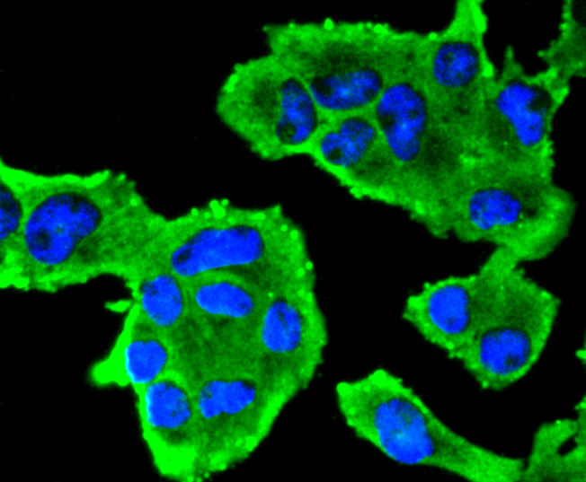
Fig4: ICC staining Insulin Receptor in RH-35 cells (green). The nuclear counter stain is DAPI (blue). Cells were fixed in paraformaldehyde, permeabilised with 0.25% Triton X100/PBS.Application
Fig5: ICC staining Insulin Receptor in PANC-1 cells (green). The nuclear counter stain is DAPI (blue). Cells were fixed in paraformaldehyde, permeabilised with 0.25% Triton X100/PBS.| Product Name | Insulin Receptor Alpha [SN20-13] |
|---|---|
| Antibody Type | Primary Antibodies |
| Antigen Alias | CD220 antibody HHF5 antibody HIR A antibody INSR antibody Insulin receptor antibody Insulin receptor subunit alpha antibody IR antibody |
| Product description | The insulin receptor (IR) is a heterodimeric protein complex that has an intracellular β subunit and an extracellular α subunit, which is disulfide- linked to a transmembrane segment. The insulin ligand binds to the IR and initiates molecular signaling pathways that promote glucose uptake in cells and glycogen synthesis. Insulin binding to IR induces phosphorylation of intracellular tyrosine kinase domains and recruitment of multiple SH2 and SH3 domain-containing intracellular proteins that serve as signaling intermediates for pleiotropic effects of insulin. The human insulin receptor gene maps to chromosome 19p13.3-p13.2 and encodes a 1382 amino acid protein that cleaves to form α and β subunits. Type 1 diabetes is an auto-immune condition of the endocrine pancreas that results in destruction of insulin secreting cells and a progressive loss in insulin-sensitive glucose uptake by cells. Type 2 diabetes is a condition where cells become resistant to insulin action. |
| Immunogen | recombinant protein |
| Clonality | Monoclonal |
|---|---|
| Isotype | IgG |
| Host Species | Recombinant rabbit |
| Tested Applications | WBICC/IF |
| WB:1:1,000-1:2,000 ICC:1:100-1:500 |
|
| Species Reactivity | HumanMouseRat |
| Concentration | 1mg/ml |
| Alternative Names | CD220 antibody HHF5 antibody HIR A antibody INSR antibody Insulin receptor antibody Insulin receptor subunit alpha antibody IR antibody |
|---|---|
| Molecular Weight(MW) | 95 kDa |
| Cellular Localization | Cell membrane. |
Application
Fig1: Western blot analysis of Insulin Receptor on different lysates using anti-Insulin Receptor antibody at 1/1,000 dilution. Positive control: Lane 1: 293 Lane 2: MCF-7 Lane 3: Hela
Application
Fig2: ICC staining Insulin Receptor in HepG2 cells (green). The nuclear counter stain is DAPI (blue). Cells were fixed in paraformaldehyde, permeabilised with 0.25% Triton X100/PBS.
Application
Fig3: ICC staining Insulin Receptor in LO2 cells (green). The nuclear counter stain is DAPI (blue). Cells were fixed in paraformaldehyde, permeabilised with 0.25% Triton X100/PBS.
Application
Fig4: ICC staining Insulin Receptor in RH-35 cells (green). The nuclear counter stain is DAPI (blue). Cells were fixed in paraformaldehyde, permeabilised with 0.25% Triton X100/PBS.
Application
Fig5: ICC staining Insulin Receptor in PANC-1 cells (green). The nuclear counter stain is DAPI (blue). Cells were fixed in paraformaldehyde, permeabilised with 0.25% Triton X100/PBS.| Positive Control | 293, MCF-7, Hela, HepG2, LO2, RH-35, PANC-1. |
|---|---|
| Application Notes | WB:1:1,000-1:2,000 ICC:1:100-1:500 |
| Form | Liquid |
|---|---|
| Storage Instructions | Store at +4℃ after thawing. Aliquot store at -20℃ or -80℃. Avoid repeated freeze / thaw cycles. |
| Storage Buffer | 1*TBS (pH7.4), 1%BSA, 40%Glycerol. Preservative: 0.05% Sodium Azide. |


