Application
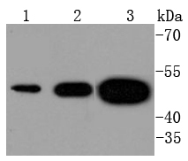
Fig1: Western blot analysis of Tissue Factor on different lysates using anti-Tissue Factor antibody at 1/1,000 dilution. Positive control: Lane 1: U937 Lane 2: SH-SY-5Y Lane 3: Mouse brainApplication
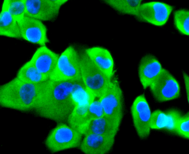
Fig2: ICC staining Tissue Factor in PANC-1 cells (green). The nuclear counter stain is DAPI (blue). Cells were fixed in paraformaldehyde, permeabilised with 0.25% Triton X100/PBS.Application
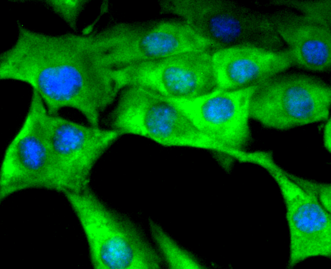
Fig3: ICC staining Tissue Factor in SHG-44 cells (green). The nuclear counter stain is DAPI (blue). Cells were fixed in paraformaldehyde, permeabilised with 0.25% Triton X100/PBS.Application
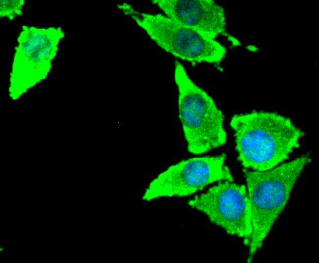
Fig4: ICC staining Tissue Factor in SH-SY-5Y cells (green). The nuclear counter stain is DAPI (blue). Cells were fixed in paraformaldehyde, permeabilised with 0.25% Triton X100/PBS.Application
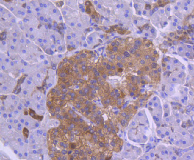
Fig5: Immunohistochemical analysis of paraffin-embedded human pancreas tissue using anti-Tissue Factor antibody. Counter stained with hematoxylin.Application
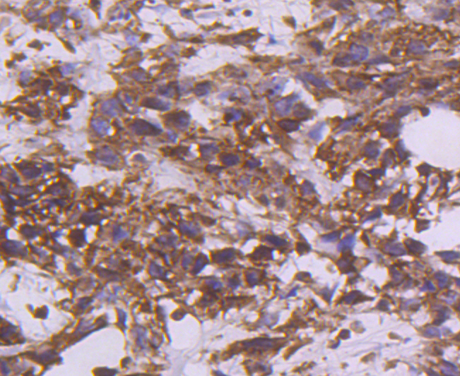
Fig6: Immunohistochemical analysis of paraffin-embedded human breast carcinoma tissue using anti-Tissue Factor antibody. Counter stained with hematoxylin.Application
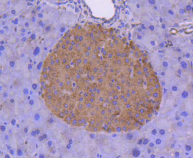
Fig7: Immunohistochemical analysis of paraffin-embedded mouse pancreas tissue using anti-Tissue Factor antibody. Counter stained with hematoxylin.| Product Name | Tissue Factor [SN20-16] |
|---|---|
| Antibody Type | Primary Antibodies |
| Antigen Alias | CD142 antibody CD142 antigen antibody Coagulation factor III (thromboplastin tissue factor) antibody Coagulation factor III antibody F3 antibody FLJ17960 antibody TF antibody TF_HUMAN antibody TFA antibody Thromboplastin antibody Tissue factor antibody |
| Product description | Hemostasis following tissue injury involves the deployment of essential plasma procoagulants (Prothrombin and Factors X, IX, V and VIII), which are involved in a blood coagulation cascade leading to the formation of insoluble Fibrin clots and the promotion of platelet aggregation. Coagulation Factor V (Factor V, FV, proaccelerin, labile factor) is a 2196 amino acid, single chain glycoprotein that is cleaved by Thrombin to yield an active, Ca2+-dependent dimer that is essential to the blood coagulation cascade. Together with catalytic Factor Xa and Ca2+ on the surface of platelets or endothelial cells, Factor Va coordinates into a Prothrombinase complex, which mediates proteolysis of Prothrombin into active Thrombin. Tissue factor (TF), also designated coagulation Factor III is a cell surface glycoprotein that enables cells to initiate blood coagulation cascades. It functions as a high-affinity receptor for coagulation Factor VII. |
| Immunogen | recombinant protein |
| Clonality | Monoclonal |
|---|---|
| Isotype | IgG |
| Host Species | Recombinant rabbit |
| Tested Applications | WBICC/IFIHC |
| WB:1:1,000-1:5,000 ICC:1:100-1:500 IHC:1:100-1:500 |
|
| Species Reactivity | HumanMouseRat |
| Concentration | 1mg/ml |
| Alternative Names | CD142 antibody CD142 antigen antibody Coagulation factor III (thromboplastin tissue factor) antibody Coagulation factor III antibody F3 antibody FLJ17960 antibody TF antibody TF_HUMAN antibody TFA antibody Thromboplastin antibody Tissue factor antibody |
|---|---|
| Molecular Weight(MW) | 50 kDa |
| Cellular Localization | Membrane,screted. |
Application
Fig1: Western blot analysis of Tissue Factor on different lysates using anti-Tissue Factor antibody at 1/1,000 dilution. Positive control: Lane 1: U937 Lane 2: SH-SY-5Y Lane 3: Mouse brain
Application
Fig2: ICC staining Tissue Factor in PANC-1 cells (green). The nuclear counter stain is DAPI (blue). Cells were fixed in paraformaldehyde, permeabilised with 0.25% Triton X100/PBS.
Application
Fig3: ICC staining Tissue Factor in SHG-44 cells (green). The nuclear counter stain is DAPI (blue). Cells were fixed in paraformaldehyde, permeabilised with 0.25% Triton X100/PBS.
Application
Fig4: ICC staining Tissue Factor in SH-SY-5Y cells (green). The nuclear counter stain is DAPI (blue). Cells were fixed in paraformaldehyde, permeabilised with 0.25% Triton X100/PBS.
Application
Fig5: Immunohistochemical analysis of paraffin-embedded human pancreas tissue using anti-Tissue Factor antibody. Counter stained with hematoxylin.
Application
Fig6: Immunohistochemical analysis of paraffin-embedded human breast carcinoma tissue using anti-Tissue Factor antibody. Counter stained with hematoxylin.
Application
Fig7: Immunohistochemical analysis of paraffin-embedded mouse pancreas tissue using anti-Tissue Factor antibody. Counter stained with hematoxylin.| Positive Control | PANC-1, SHG-44, SH-SY-5Y, U937, human breast carcinoma tissue, human pancreas tissue, mouse pancreas tissue. |
|---|---|
| Application Notes | WB:1:1,000-1:5,000 ICC:1:100-1:500 IHC:1:100-1:500 |
| Form | Liquid |
|---|---|
| Storage Instructions | Store at +4℃ after thawing. Aliquot store at -20℃ or -80℃. Avoid repeated freeze / thaw cycles. |
| Storage Buffer | 1*TBS (pH7.4), 1%BSA, 40%Glycerol. Preservative: 0.05% Sodium Azide. |


