Application
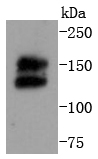
Fig1: Western blot analysis of Ret on MCF-7 cells lysates using anti-Ret antibody at 1/1,000 dilution.Application
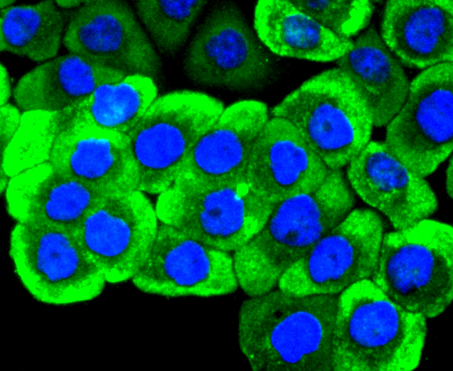
Fig2: ICC staining Ret in AGS cells (green). The nuclear counter stain is DAPI (blue). Cells were fixed in paraformaldehyde, permeabilised with 0.25% Triton X100/PBS.Application
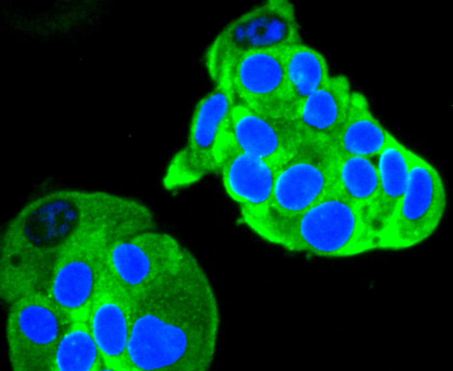
Fig3: ICC staining Ret in MCF-7 cells (green). The nuclear counter stain is DAPI (blue). Cells were fixed in paraformaldehyde, permeabilised with 0.25% Triton X100/PBS.Application
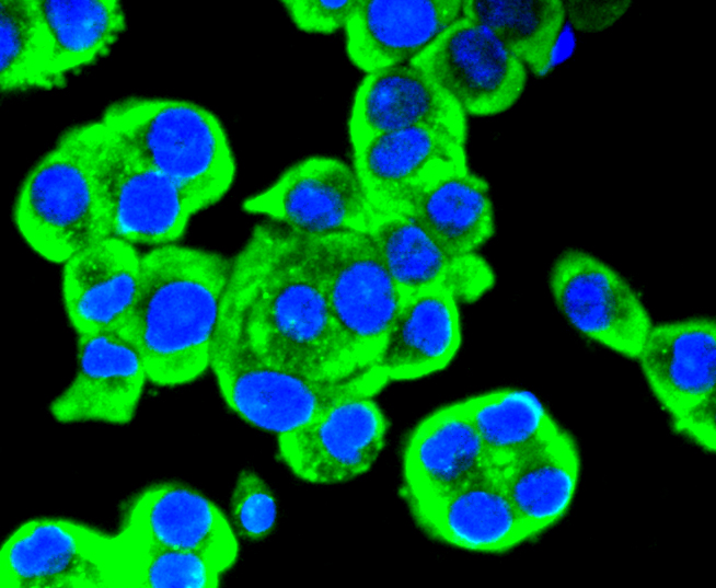
Fig4: ICC staining Ret in SW480 cells (green). The nuclear counter stain is DAPI (blue). Cells were fixed in paraformaldehyde, permeabilised with 0.25% Triton X100/PBS.Application
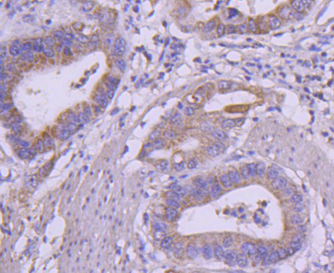
Fig5: Immunohistochemical analysis of paraffin-embedded human gastric carcinoma tissue using anti-Ret antibody. Counter stained with hematoxylin.Application
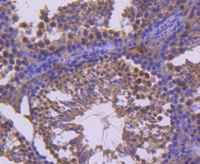
Fig6: Immunohistochemical analysis of paraffin-embedded mouse testis tissue using anti-Ret antibody. Counter stained with hematoxylin.Application
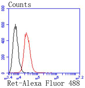
Fig7: Flow cytometric analysis of SW480 cells with Ret antibody at 1/50 dilution (red) compared with an unlabelled control (cells without incubation with primary antibody; black). Alexa Fluor 488-conjugated goat anti rabbit IgG was used as the secondary antibody.| Product Name | Ret [SN20-28] |
|---|---|
| Antibody Type | Primary Antibodies |
| Antigen Alias | C ret antibody Cadherin family member 12 antibody Cadherin related family member 16 antibody CDHF 12 antibody CDHF12 antibody CDHR16 antibody ELKS Fusion gene antibody HSCR 1 antibody HSCR1 antibody Hydroxyaryl protein kinase antibody MEN2A antibody MEN2B antibody MTC 1 antibody MTC1 antibody Multiple endocrine neoplasia and medullary thyroid carcinoma 1 antibody Oncogene RET antibody Proto oncogene tyrosine protein kinase receptor ret antibody Proto-oncogene c-Ret antibody Proto-oncogene tyrosine-protein kinase receptor ret antibody PTC antibody RET antibody RET ELE1 antibody Ret Proto oncogene antibody RET transforming sequence antibody RET_HUMAN antibody RET51 antibody RET9 antibody tyrosine-protein kinase receptor ret antibody |
| Product description | The Ret proto-oncogene (c-Ret) is a receptor tyrosine kinase that functions as a multicomponent receptor complex in conjunction with other membrane-bound, ligand-binding GDNF family receptors. Ligands that bind the Ret receptor include the glial cell line-derived neurotrophic factor (GDNF) and its congeners neurturin, persephin, and artemin. Research studies have shown that alterations in the corresponding RET gene are associated with diseases including papillary thyroid carcinoma, multiple endocrine neoplasia (type 2A and 2B), familial medullary thyroid carcinoma, and a congenital developmental disorder known as Hirschsprung’s disease. The Tyr905 residue located in the Ret kinase domain plays a crucial role in Ret catalytic and biological activity. Substitution of Phe for Tyr at position 905 dramatically inhibits Ret autophosphorylation activity. |
| Immunogen | recombinant protein |
| Clonality | Monoclonal |
|---|---|
| Isotype | IgG |
| Host Species | Recombinant rabbit |
| Tested Applications | WBICC/IFIHCIPFC |
| WB:1:1,000-1:2,000 ICC:1:100-1:500 IHC:1:50-1:200 FC:1:50-1:100 |
|
| Species Reactivity | HumanMouseRat |
| Concentration | 1mg/ml |
| Alternative Names | C ret antibody Cadherin family member 12 antibody Cadherin related family member 16 antibody CDHF 12 antibody CDHF12 antibody CDHR16 antibody ELKS Fusion gene antibody HSCR 1 antibody HSCR1 antibody Hydroxyaryl protein kinase antibody MEN2A antibody MEN2B antibody MTC 1 antibody MTC1 antibody Multiple endocrine neoplasia and medullary thyroid carcinoma 1 antibody Oncogene RET antibody Proto oncogene tyrosine protein kinase receptor ret antibody Proto-oncogene c-Ret antibody Proto-oncogene tyrosine-protein kinase receptor ret antibody PTC antibody RET antibody RET ELE1 antibody Ret Proto oncogene antibody RET transforming sequence antibody RET_HUMAN antibody RET51 antibody RET9 antibody tyrosine-protein kinase receptor ret antibody |
|---|---|
| Molecular Weight(MW) | 150/175 kDa |
| Cellular Localization | Cell membrane, Endosome membrane. |
Application
Fig1: Western blot analysis of Ret on MCF-7 cells lysates using anti-Ret antibody at 1/1,000 dilution.
Application
Fig2: ICC staining Ret in AGS cells (green). The nuclear counter stain is DAPI (blue). Cells were fixed in paraformaldehyde, permeabilised with 0.25% Triton X100/PBS.
Application
Fig3: ICC staining Ret in MCF-7 cells (green). The nuclear counter stain is DAPI (blue). Cells were fixed in paraformaldehyde, permeabilised with 0.25% Triton X100/PBS.
Application
Fig4: ICC staining Ret in SW480 cells (green). The nuclear counter stain is DAPI (blue). Cells were fixed in paraformaldehyde, permeabilised with 0.25% Triton X100/PBS.
Application
Fig5: Immunohistochemical analysis of paraffin-embedded human gastric carcinoma tissue using anti-Ret antibody. Counter stained with hematoxylin.
Application
Fig6: Immunohistochemical analysis of paraffin-embedded mouse testis tissue using anti-Ret antibody. Counter stained with hematoxylin.
Application
Fig7: Flow cytometric analysis of SW480 cells with Ret antibody at 1/50 dilution (red) compared with an unlabelled control (cells without incubation with primary antibody; black). Alexa Fluor 488-conjugated goat anti rabbit IgG was used as the secondary antibody.| Positive Control | AGS, MCF-7, SW480, mouse testis tissue, human gastric carcinoma tissue. |
|---|---|
| Application Notes | WB:1:1,000-1:2,000 ICC:1:100-1:500 IHC:1:50-1:200 FC:1:50-1:100 |
| Form | Liquid |
|---|---|
| Storage Instructions | Store at +4℃ after thawing. Aliquot store at -20℃ or -80℃. Avoid repeated freeze / thaw cycles. |
| Storage Buffer | 1*TBS (pH7.4), 1%BSA, 40%Glycerol. Preservative: 0.05% Sodium Azide. |


