WB
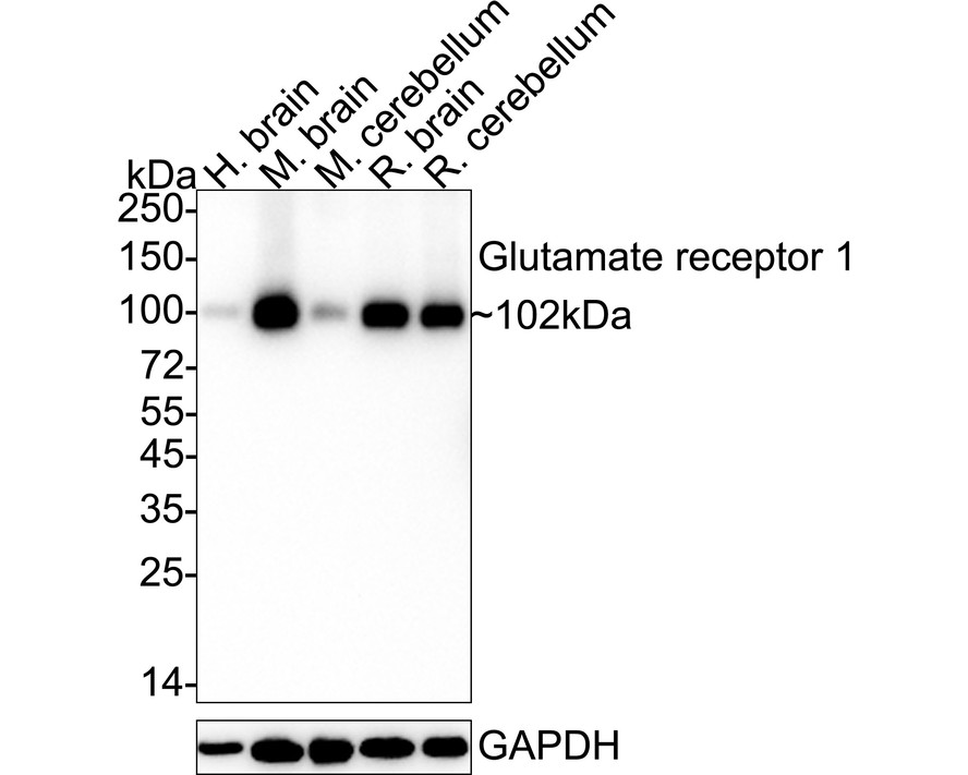
Western blot analysis of Glutamate receptor 1 on different lysates with Rabbit anti-Glutamate receptor 1 antibody at 1/1,000 dilution. Lane 1: Human brain tissue lysate, Lane 2: Mouse brain tissue lysate, Lane 3: Mouse cerebellum tissue lysate, Lane 4: Rat brain tissue lysate, Lane 5: Rat cerebellum tissue lysate, Lysates/proteins at 40 µg/Lane. Exposure time: 2 minutes; 4-20% SDS-PAGE gel. Proteins were transferred to a PVDF membrane and blocked with 5% NFDM/TBST for 1 hour at room temperature. The primary antibody at 1/1,000 dilution was used in 5% NFDM/TBST at 4℃ overnight. Goat Anti-Rabbit IgG - HRP Secondary Antibody at 1/50,000 dilution was used for 1 hour at room temperature.IHC
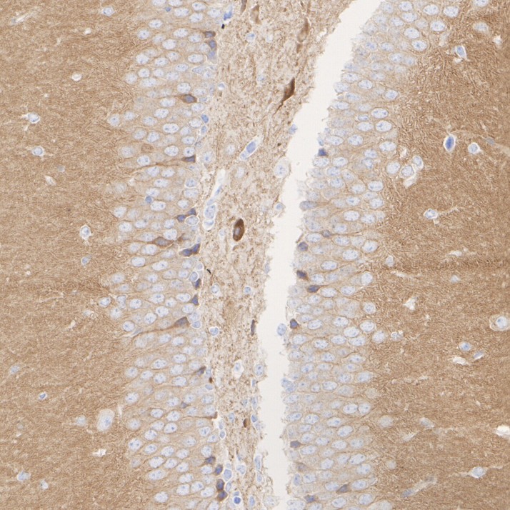
Immunohistochemical analysis of paraffin-embedded mouse hippocampus tissue with Rabbit anti-Glutamate receptor 1 antibody at 1/1,000 dilution. The section was pre-treated using heat mediated antigen retrieval with Tris-EDTA buffer (pH 9.0) for 20 minutes. The tissues were blocked in 1% BSA for 20 minutes at room temperature, washed with ddH2O and PBS, and then probed with the primary antibody at 1/1,000 dilution for 1 hour at room temperature. The detection was performed using an HRP conjugated compact polymer system. DAB was used as the chromogen. Tissues were counterstained with hematoxylin and mounted with DPX.IHC
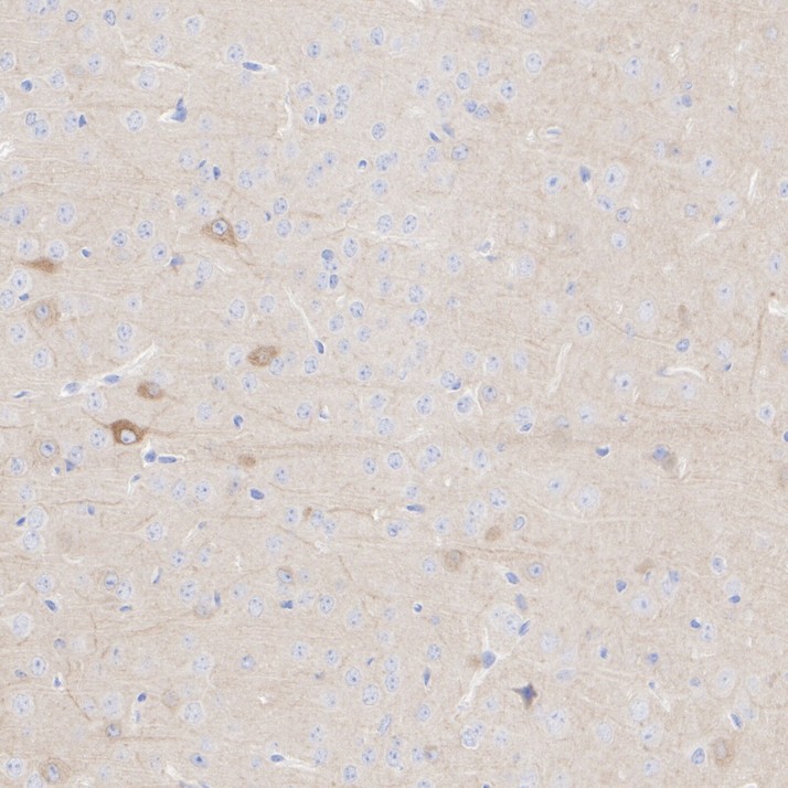
Immunohistochemical analysis of paraffin-embedded mouse cerebral cortex tissue with Rabbit anti-Glutamate receptor 1 antibody at 1/1,000 dilution. The section was pre-treated using heat mediated antigen retrieval with Tris-EDTA buffer (pH 9.0) for 20 minutes. The tissues were blocked in 1% BSA for 20 minutes at room temperature, washed with ddH2O and PBS, and then probed with the primary antibody at 1/1,000 dilution for 1 hour at room temperature. The detection was performed using an HRP conjugated compact polymer system. DAB was used as the chromogen. Tissues were counterstained with hematoxylin and mounted with DPX.IHC
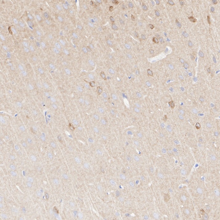
Immunohistochemical analysis of paraffin-embedded rat cerebral cortex tissue with Rabbit anti-Glutamate receptor 1 antibody at 1/1,000 dilution. The section was pre-treated using heat mediated antigen retrieval with Tris-EDTA buffer (pH 9.0) for 20 minutes. The tissues were blocked in 1% BSA for 20 minutes at room temperature, washed with ddH2O and PBS, and then probed with the primary antibody at 1/1,000 dilution for 1 hour at room temperature. The detection was performed using an HRP conjugated compact polymer system. DAB was used as the chromogen. Tissues were counterstained with hematoxylin and mounted with DPX.IF-F
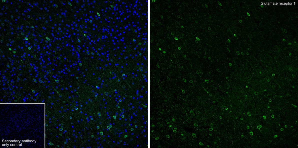
Immunofluorescence analysis of frozen mouse brain tissue with Rabbit anti-Glutamate receptor 1 antibody at 1/200 dilution. The section was pre-treated using heat mediated antigen retrieval with sodium citrate buffer (pH 6.0) for about 2 minutes in microwave oven. The tissues were blocked in 10% negative goat serum for 1 hour at room temperature, washed with PBS, and then probed with the primary antibody (green) at 1/200 dilution overnight at 4 ℃, washed with PBS. Goat Anti-Rabbit IgG H&L (488) was used as the secondary antibody at 1/1,000 dilution. Nuclei were counterstained with DAPI (blue).| Product Name | Glutamate receptor 1 Recombinant Rabbit Monoclonal Antibody |
|---|---|
| Antibody Type | Primary Antibodies |
| Product description | Glutamate receptors mediate most excitatory neurotransmission in the brain and play an important role in neural plasticity, neural development and neurodegeneration. Ionotropic glutamate receptors are categorized into NMDA receptors and kainate/AMPA receptors, both of which contain glutamate-gated, cation-specific ion channels. Kainate/AMPA receptors are co-localized with NMDA receptors in many synapses and consist of seven structurally related subunits designated GluR-1 to -7. The kainate/AMPA receptors are primarily responsible for the fast excitatory neuro-transmission by glutamate whereas the NMDA receptors are functionally characterized by a slow kinetic and a high permeability for Ca2+ ions. The NMDA receptors consist of five subunits: epsilion 1, 2, 3, 4 and one zeta subunit. The zeta subunit is expressed throughout the brainstem whereas the four epsilon subunits display limited distribution. |
| Immunogen | Synthetic peptide within Human Glutamate receptor 1 aa 857-906 / 906. |
| Clonality | Monoclonal |
|---|---|
| Isotype | IgG |
| Host Species | Recombinant rabbit |
| Tested Applications | IF-FIHCWB |
| WB:1:1000 IHC:1:500-1:1000 IF-F:1:200-1:500 |
|
| Species Reactivity | HumanMouseRat |
| Concentration | 1mg/ml |
| Purification | Protein A |
| Alternative Names | GLUR 1 antibody
GLUR A antibody
AMPA 1 antibody
AMPA selective glutamate receptor 1 antibody
AMPA-selective glutamate receptor 1 antibody
GluA1 antibody
GLUH1 antibody
GluR K1 antibody
GluR-1 antibody
GluR-A antibody
GluR-K1 antibody
GLUR1 antibody
GLURA antibody
GluRK1 antibody
Glutamate receptor 1 antibody
Glutamate receptor ionotropic AMPA 1 antibody
Glutamate receptor ionotropic antibody
Glutamate receptor ionotropic AMPA 1 antibody Gria1 antibody GRIA1_HUMAN antibody HBGR1 antibody MGC133252 antibody OTTHUMP00000160643 antibody OTTHUMP00000165781 antibody OTTHUMP00000224241 antibody OTTHUMP00000224242 antibody OTTHUMP00000224243 antibody |
|---|---|
| Molecular Weight(MW) | 102 kDa |
| Cellular Localization | Cell membrane, postsynaptic cell membrane, postsynaptic density membrane, Endoplasmic reticulum membrane, Early endosome membrane, Recycling endosome membrane, dendrite, dendritic spine. |
WB
Western blot analysis of Glutamate receptor 1 on different lysates with Rabbit anti-Glutamate receptor 1 antibody at 1/1,000 dilution. Lane 1: Human brain tissue lysate, Lane 2: Mouse brain tissue lysate, Lane 3: Mouse cerebellum tissue lysate, Lane 4: Rat brain tissue lysate, Lane 5: Rat cerebellum tissue lysate, Lysates/proteins at 40 µg/Lane. Exposure time: 2 minutes; 4-20% SDS-PAGE gel. Proteins were transferred to a PVDF membrane and blocked with 5% NFDM/TBST for 1 hour at room temperature. The primary antibody at 1/1,000 dilution was used in 5% NFDM/TBST at 4℃ overnight. Goat Anti-Rabbit IgG - HRP Secondary Antibody at 1/50,000 dilution was used for 1 hour at room temperature.
IHC
Immunohistochemical analysis of paraffin-embedded mouse hippocampus tissue with Rabbit anti-Glutamate receptor 1 antibody at 1/1,000 dilution. The section was pre-treated using heat mediated antigen retrieval with Tris-EDTA buffer (pH 9.0) for 20 minutes. The tissues were blocked in 1% BSA for 20 minutes at room temperature, washed with ddH2O and PBS, and then probed with the primary antibody at 1/1,000 dilution for 1 hour at room temperature. The detection was performed using an HRP conjugated compact polymer system. DAB was used as the chromogen. Tissues were counterstained with hematoxylin and mounted with DPX.
IHC
Immunohistochemical analysis of paraffin-embedded mouse cerebral cortex tissue with Rabbit anti-Glutamate receptor 1 antibody at 1/1,000 dilution. The section was pre-treated using heat mediated antigen retrieval with Tris-EDTA buffer (pH 9.0) for 20 minutes. The tissues were blocked in 1% BSA for 20 minutes at room temperature, washed with ddH2O and PBS, and then probed with the primary antibody at 1/1,000 dilution for 1 hour at room temperature. The detection was performed using an HRP conjugated compact polymer system. DAB was used as the chromogen. Tissues were counterstained with hematoxylin and mounted with DPX.
IHC
Immunohistochemical analysis of paraffin-embedded rat cerebral cortex tissue with Rabbit anti-Glutamate receptor 1 antibody at 1/1,000 dilution. The section was pre-treated using heat mediated antigen retrieval with Tris-EDTA buffer (pH 9.0) for 20 minutes. The tissues were blocked in 1% BSA for 20 minutes at room temperature, washed with ddH2O and PBS, and then probed with the primary antibody at 1/1,000 dilution for 1 hour at room temperature. The detection was performed using an HRP conjugated compact polymer system. DAB was used as the chromogen. Tissues were counterstained with hematoxylin and mounted with DPX.
IF-F
Immunofluorescence analysis of frozen mouse brain tissue with Rabbit anti-Glutamate receptor 1 antibody at 1/200 dilution. The section was pre-treated using heat mediated antigen retrieval with sodium citrate buffer (pH 6.0) for about 2 minutes in microwave oven. The tissues were blocked in 10% negative goat serum for 1 hour at room temperature, washed with PBS, and then probed with the primary antibody (green) at 1/200 dilution overnight at 4 ℃, washed with PBS. Goat Anti-Rabbit IgG H&L (488) was used as the secondary antibody at 1/1,000 dilution. Nuclei were counterstained with DAPI (blue).| Application Notes | WB:1:1000 IHC:1:500-1:1000 IF-F:1:200-1:500 |
|---|
| Form | Liquid |
|---|---|
| Storage Instructions | Store at +4℃ after thawing. Aliquot store at -20℃ or -80℃. Avoid repeated freeze / thaw cycles. |
| Storage Buffer | 1*TBS (pH7.4), 1%BSA, 40%Glycerol. Preservative: 0.05% Sodium Azide. |


