WB
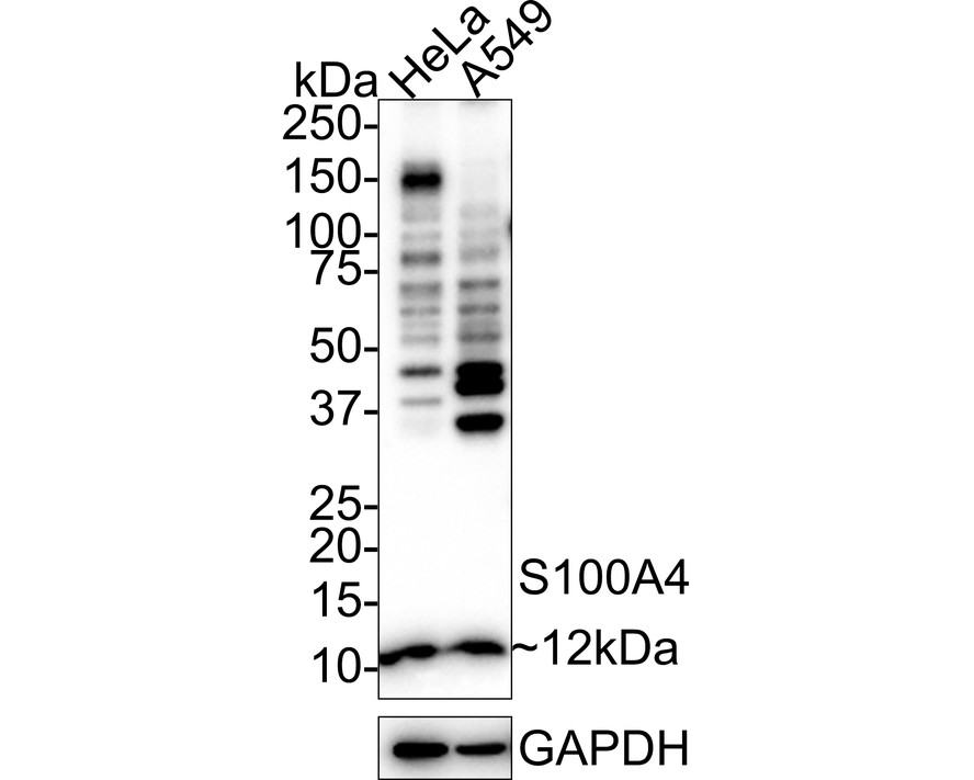
Western blot analysis of S100A4 on different lysates with Rabbit anti-S100A4 antibody at 1/1,000 dilution. Lane 1: HeLa cell lysate Lane 2: A549 cell lysate Lysates/proteins at 20 µg/Lane. Exposure time: 3 minutes; 4-20% SDS-PAGE gel. Proteins were transferred to a PVDF membrane and blocked with 5% NFDM/TBST for 1 hour at room temperature. The primary antibody at 1/1,000 dilution was used in 5% NFDM/TBST at 4℃ overnight. Goat Anti-Rabbit IgG - HRP Secondary Antibody at 1/50,000 dilution was used for 1 hour at room temperature.WB
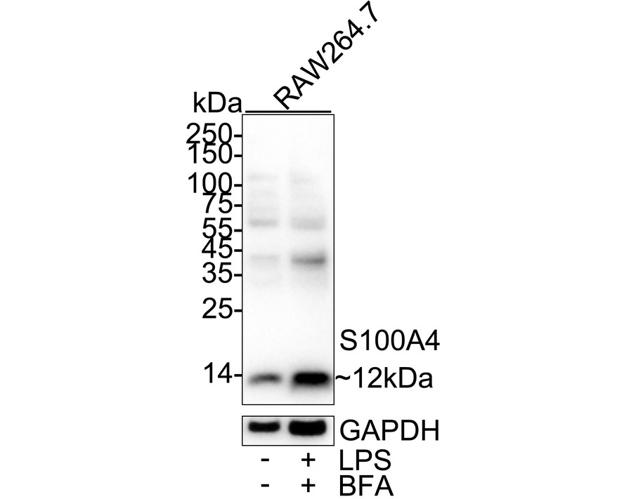
Western blot analysis of S100A4 on different lysates with Rabbit anti-S100A4 antibody at 1/1,000 dilution. Lane 1: RAW264.7 cell lysate, Lane 2: RAW264.7 treated with 100ng/mL LPS for 7 hours add 300ng/mL BFA for last 3 hours cell lysate, Lysates/proteins at 20 µg/Lane. Exposure time: 20 seconds; 4-20% SDS-PAGE gel. Proteins were transferred to a PVDF membrane and blocked with 5% NFDM/TBST for 1 hour at room temperature. The primary antibody at 1/1,000 dilution was used in primary antibody dilution at 4℃ overnight. Goat Anti-Rabbit IgG - HRP Secondary Antibody at 1/50,000 dilution was used for 1 hour at room temperature.IHC
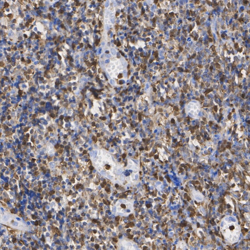
Immunohistochemical analysis of paraffin-embedded human tonsil tissue with Rabbit anti-S100A4 antibody at 1/1,000 dilution. The section was pre-treated using heat mediated antigen retrieval with Tris-EDTA buffer (pH 9.0) for 20 minutes. The tissues were blocked in 1% BSA for 20 minutes at room temperature, washed with ddH2O and PBS, and then probed with the primary antibody at 1/1,000 dilution for 1 hour at room temperature. The detection was performed using an HRP conjugated compact polymer system. DAB was used as the chromogen. Tissues were counterstained with hematoxylin and mounted with DPX.IHC
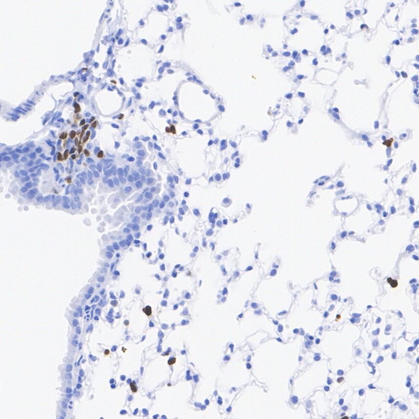
Immunohistochemical analysis of paraffin-embedded mouse lung tissue with Rabbit anti-S100A4 antibody at 1/1,000 dilution. The section was pre-treated using heat mediated antigen retrieval with Tris-EDTA buffer (pH 9.0) for 20 minutes. The tissues were blocked in 1% BSA for 20 minutes at room temperature, washed with ddH2O and PBS, and then probed with the primary antibody at 1/1,000 dilution for 1 hour at room temperature. The detection was performed using an HRP conjugated compact polymer system. DAB was used as the chromogen. Tissues were counterstained with hematoxylin and mounted with DPX.ICC/IF
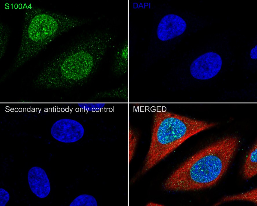
Immunocytochemistry analysis of HeLa cells labeling S100A4 with Rabbit anti-S100A4 antibody at 1/100 dilution. Cells were fixed in 4% paraformaldehyde for 15 minutes at room temperature, permeabilized with 0.1% Triton X-100 in PBS for 15 minutes at room temperature, then blocked with 1% BSA in 10% negative goat serum for 1 hour at room temperature. Cells were then incubated with Rabbit anti-S100A4 antibody at 1/100 dilution in 1% BSA in PBST overnight at 4 ℃. Goat Anti-Rabbit IgG H&L (488) was used as the secondary antibody at 1/1,000 dilution. PBS instead of the primary antibody was used as the secondary antibody only control. Nuclear DNA was labelled in blue with DAPI Beta tubulin (red) was stained at 1/100 dilution overnight at +4℃. Goat Anti-Mouse IgG H&L (594) was used as the secondary antibody at 1/1,000 dilution.FC
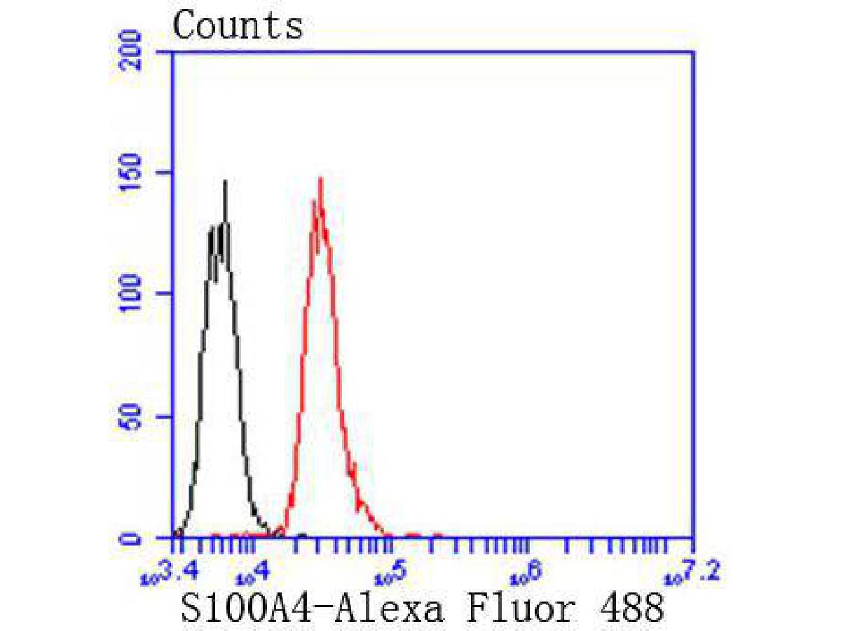
Flow cytometric analysis of S100A4 was done on Jurkat cells. The cells were fixed, permeabilized and stained with the primary antibody (1/50) (red). After incubation of the primary antibody at room temperature for an hour, the cells were stained with a Alexa Fluor®488 conjugate-Goat anti-Rabbit IgG Secondary antibody at 1/1000 dilution for 30 minutes.Unlabelled sample was used as a control (cells without incubation with primary antibody; black).| Product Name | S100A4 Recombinant Rabbit Monoclonal Antibody |
|---|---|
| Antibody Type | Primary Antibodies |
| Product description | The Mts1 gene encodes a small acidic Ca2+-binding protein, Mts1 (also designated S100A4, calvasculin or metastasin). Mts1 belongs to the S100 family of small Ca2+-binding proteins and is expressed in a cell-specific manner. Mts1 protein is involved in tumor progression and metastasis, and also has a significant stimulatory effect on angiogenesis. The level of Mts1 protein in serum increases with aging, suggesting that Mts1 may play a role in the ind-uction of tumor progression via stimulation of angiogenesis. In addition, Mts1 cooperates with p53 in apoptosis induction by binding to the C-term-inal regulatory domain of p53 to inhibit the DNA binding activity of p53. The ability of Mts1 to enhance p53-dependent apoptosis may accelerate the loss of p53 function in tumors. Thus, Mts1 can contribute to the development of a more aggressive phenotype during tumor progression. |
| Immunogen | Recombinant protein within Human S100A4 aa 1-101 / 101. |
| Clonality | Monoclonal |
|---|---|
| Isotype | IgG |
| Host Species | Recombinant rabbit |
| Tested Applications | FCICC/IFIHCWB |
| WB:1:1000 IHC:1:500-1:1000 ICC/IF:1:100-1:200 FC:1:50-1:100 |
|
| Species Reactivity | HumanMouseRat |
| Concentration | 1mg/ml |
| Purification | Protein A |
| Alternative Names | 18A2 antibody
42A antibody
calcium Placental protein antibody
Calvasculin antibody
CAPL antibody
Fibroblast specific protein 1 (FSP1) antibody
Fibroblast specific protein 1 antibody
Fibroblast specific protein antibody
FSP1 antibody
Leukemia multidrug resistance associated protein antibody
Malignant transformation suppression 1 (MTS1) antibody
Malignant transformation suppression 1 antibody
Metastasin antibody
MTS1 antibody
OTTHUMP00000015467 antibody
OTTHUMP00000015468 antibody
P9KA antibody
PEL98 antibody
Placental calcium-binding protein antibody
Protein Mts1 antibody
Protein S100 A4 antibody
Protein S100-A4 antibody
S100 calcium binding protein A4 (calcium protein calvasculin metastasin murine placental homolog) antibody S100 calcium binding protein A4 antibody S100 calcium-binding protein A4 antibody S100a4 antibody S10A4_HUMAN antibody |
|---|---|
| Molecular Weight(MW) | 12 kDa |
| Cellular Localization | Extracellular space, nucleus |
WB
Western blot analysis of S100A4 on different lysates with Rabbit anti-S100A4 antibody at 1/1,000 dilution. Lane 1: HeLa cell lysate Lane 2: A549 cell lysate Lysates/proteins at 20 µg/Lane. Exposure time: 3 minutes; 4-20% SDS-PAGE gel. Proteins were transferred to a PVDF membrane and blocked with 5% NFDM/TBST for 1 hour at room temperature. The primary antibody at 1/1,000 dilution was used in 5% NFDM/TBST at 4℃ overnight. Goat Anti-Rabbit IgG - HRP Secondary Antibody at 1/50,000 dilution was used for 1 hour at room temperature.
WB
Western blot analysis of S100A4 on different lysates with Rabbit anti-S100A4 antibody at 1/1,000 dilution. Lane 1: RAW264.7 cell lysate, Lane 2: RAW264.7 treated with 100ng/mL LPS for 7 hours add 300ng/mL BFA for last 3 hours cell lysate, Lysates/proteins at 20 µg/Lane. Exposure time: 20 seconds; 4-20% SDS-PAGE gel. Proteins were transferred to a PVDF membrane and blocked with 5% NFDM/TBST for 1 hour at room temperature. The primary antibody at 1/1,000 dilution was used in primary antibody dilution at 4℃ overnight. Goat Anti-Rabbit IgG - HRP Secondary Antibody at 1/50,000 dilution was used for 1 hour at room temperature.
IHC
Immunohistochemical analysis of paraffin-embedded human tonsil tissue with Rabbit anti-S100A4 antibody at 1/1,000 dilution. The section was pre-treated using heat mediated antigen retrieval with Tris-EDTA buffer (pH 9.0) for 20 minutes. The tissues were blocked in 1% BSA for 20 minutes at room temperature, washed with ddH2O and PBS, and then probed with the primary antibody at 1/1,000 dilution for 1 hour at room temperature. The detection was performed using an HRP conjugated compact polymer system. DAB was used as the chromogen. Tissues were counterstained with hematoxylin and mounted with DPX.
IHC
Immunohistochemical analysis of paraffin-embedded mouse lung tissue with Rabbit anti-S100A4 antibody at 1/1,000 dilution. The section was pre-treated using heat mediated antigen retrieval with Tris-EDTA buffer (pH 9.0) for 20 minutes. The tissues were blocked in 1% BSA for 20 minutes at room temperature, washed with ddH2O and PBS, and then probed with the primary antibody at 1/1,000 dilution for 1 hour at room temperature. The detection was performed using an HRP conjugated compact polymer system. DAB was used as the chromogen. Tissues were counterstained with hematoxylin and mounted with DPX.
ICC/IF
Immunocytochemistry analysis of HeLa cells labeling S100A4 with Rabbit anti-S100A4 antibody at 1/100 dilution. Cells were fixed in 4% paraformaldehyde for 15 minutes at room temperature, permeabilized with 0.1% Triton X-100 in PBS for 15 minutes at room temperature, then blocked with 1% BSA in 10% negative goat serum for 1 hour at room temperature. Cells were then incubated with Rabbit anti-S100A4 antibody at 1/100 dilution in 1% BSA in PBST overnight at 4 ℃. Goat Anti-Rabbit IgG H&L (488) was used as the secondary antibody at 1/1,000 dilution. PBS instead of the primary antibody was used as the secondary antibody only control. Nuclear DNA was labelled in blue with DAPI Beta tubulin (red) was stained at 1/100 dilution overnight at +4℃. Goat Anti-Mouse IgG H&L (594) was used as the secondary antibody at 1/1,000 dilution.
FC
Flow cytometric analysis of S100A4 was done on Jurkat cells. The cells were fixed, permeabilized and stained with the primary antibody (1/50) (red). After incubation of the primary antibody at room temperature for an hour, the cells were stained with a Alexa Fluor®488 conjugate-Goat anti-Rabbit IgG Secondary antibody at 1/1000 dilution for 30 minutes.Unlabelled sample was used as a control (cells without incubation with primary antibody; black).| Application Notes | WB:1:1000 IHC:1:500-1:1000 ICC/IF:1:100-1:200 FC:1:50-1:100 |
|---|
| Form | Liquid |
|---|---|
| Storage Instructions | Store at +4℃ after thawing. Aliquot store at -20℃ or -80℃. Avoid repeated freeze / thaw cycles. |
| Storage Buffer | 1*TBS (pH7.4), 1%BSA, 40%Glycerol. Preservative: 0.05% Sodium Azide. |


