WB
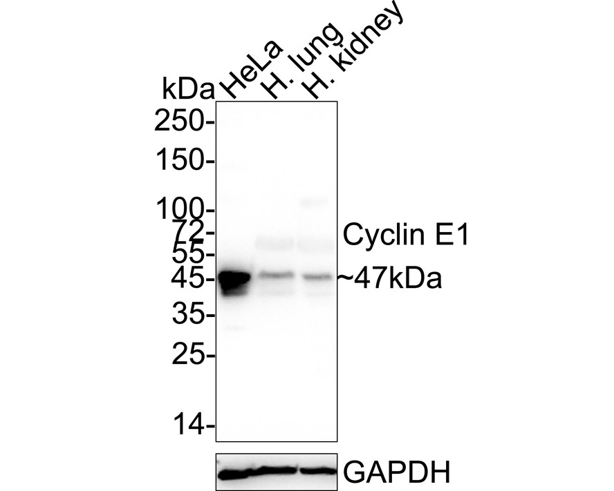
Western blot analysis of Cyclin E1 on different lysates with Rabbit anti-Cyclin E1 antibody at 1/1,000 dilution. Lane 1: HeLa cell lysate (20 µg/Lane), Lane 2: Human lung tissue lysate (40 µg/Lane), Lane 3: Human kidney tissue lysate (40 µg/Lane), Exposure time: 1 minute; 4-20% SDS-PAGE gel. Proteins were transferred to a PVDF membrane and blocked with 5% NFDM/TBST for 1 hour at room temperature. The primary antibody at 1/1,000 dilution was used in 5% NFDM/TBST at 4℃ overnight. Goat Anti-Rabbit IgG - HRP Secondary Antibody at 1/50,000 dilution was used for 1 hour at room temperature.IHC
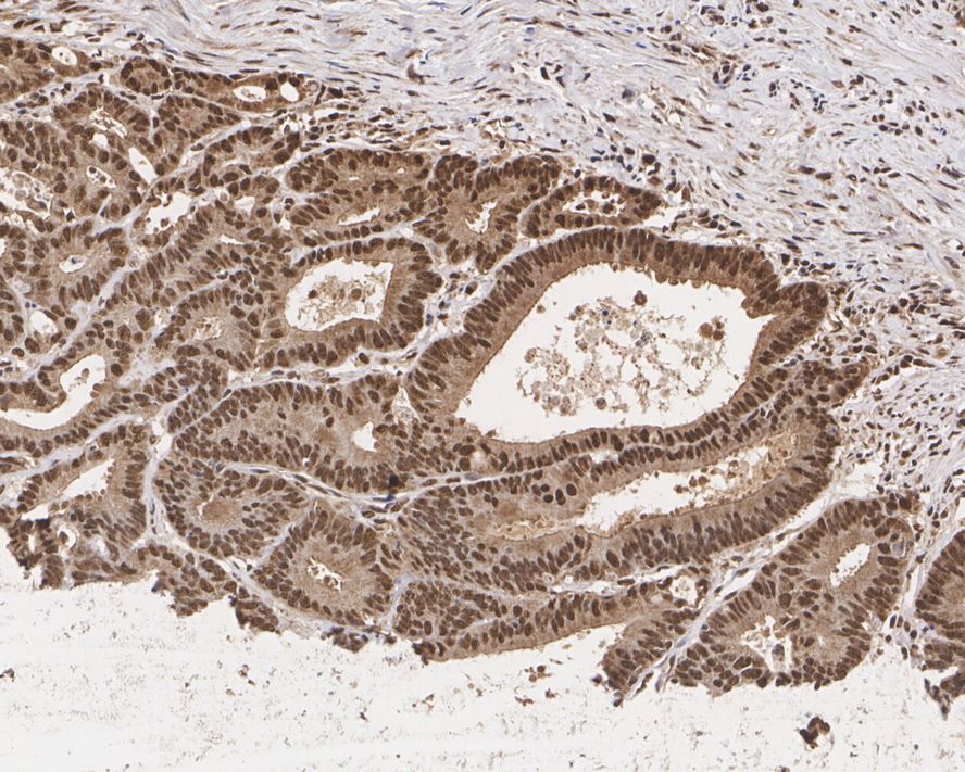
Immunohistochemical analysis of paraffin-embedded human colon carcinoma tissue with Rabbit anti-Cyclin E1 antibody at 1/1,000 dilution. The section was pre-treated using heat mediated antigen retrieval with sodium citrate buffer (pH 6.0) (high pressure) for 2 minutes. The tissues were blocked in 1% BSA for 20 minutes at room temperature, washed with ddH2O and PBS, and then probed with the primary antibody at 1/1,000 dilution for 1 hour at room temperature. The detection was performed using an HRP conjugated compact polymer system. DAB was used as the chromogen. Tissues were counterstained with hematoxylin and mounted with DPX.IHC
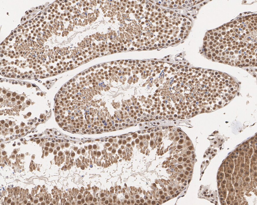
Immunohistochemical analysis of paraffin-embedded mouse testis tissue with Rabbit anti-Cyclin E1 antibody at 1/1,000 dilution. The section was pre-treated using heat mediated antigen retrieval with sodium citrate buffer (pH 6.0) (high pressure) for 2 minutes. The tissues were blocked in 1% BSA for 20 minutes at room temperature, washed with ddH2O and PBS, and then probed with the primary antibody at 1/1,000 dilution for 1 hour at room temperature. The detection was performed using an HRP conjugated compact polymer system. DAB was used as the chromogen. Tissues were counterstained with hematoxylin and mounted with DPX.ICC/IF
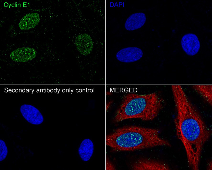
Immunocytochemistry analysis of HeLa cells labeling Cyclin E1 with Rabbit anti-Cyclin E1 antibody at 1/2,000 dilution. Cells were fixed in 4% paraformaldehyde for 15 minutes at room temperature, permeabilized with 0.1% Triton X-100 in PBS for 15 minutes at room temperature, then blocked with 1% BSA in 10% negative goat serum for 1 hour at room temperature. Cells were then incubated with Rabbit anti-Cyclin E1 antibody at 1/2,000 dilution in 1% BSA in PBST overnight at 4 ℃. Goat Anti-Rabbit IgG H&L (488) was used as the secondary antibody at 1/1,000 dilution. PBS instead of the primary antibody was used as the secondary antibody only control. Nuclear DNA was labelled in blue with DAPI. Beta tubulin (red) was stained at 1/100 dilution overnight at +4℃. Goat Anti-Mouse IgG H&L (594) was used as the secondary antibody at 1/1,000 dilution.| Product Name | Cyclin E1 Recombinant Rabbit Monoclonal Antibody |
|---|---|
| Antibody Type | Primary Antibodies |
| Product description | The protein encoded by this gene belongs to the highly conserved cyclin family, whose members are characterized by a dramatic periodicity in protein abundance through the cell cycle. Cyclins were first identified in invertebrates as proteins that oscillate dramatically through the cell cycle. These proteins have been well conserved through evolution and play a critical role in regulation of cell division. Cyclins function as regulators of CDK kinases. Different cyclins exhibit distinct expression and degradation patterns which contribute to the temporal coordination of each mitotic event. This cyclin forms a complex with and functions as a regulatory subunit of CDK2, whose activity is required for cell cycle G1/S transition. This protein accumulates at the G1-S phase boundary and is degraded as cells progress through S phase. cyclin E has been found to be associated with the transcription factor E2F in a temporally regulated manner. The cyclin E/E2F complex is detected primarily during the G1 phase of the cell cycle and decreases as cells enter S phase. Overexpression of this gene has been observed in many tumors, which results in chromosome instability, and thus may contribute to tumorigenesis. This protein was found to associate with, and be involved in, the phosphorylation of NPAT protein (nuclear protein mapped to the ATM locus), which participates in cell-cycle regulated histone gene expression and plays a critical role in promoting cell-cycle progression in the absence of pRB. |
| Immunogen | Synthetic peptide within Human Cyclin E1 aa 361-410 / 410. |
| Clonality | Monoclonal |
|---|---|
| Isotype | IgG |
| Host Species | Recombinant rabbit |
| Tested Applications | ICC/IFIHCWB |
| WB:1:500-1:1000 IHC:1:200-1:1000 ICC/IF:1:2000 |
|
| Species Reactivity | HumanMouse |
| Concentration | 1mg/ml |
| Purification | Protein A |
| Alternative Names | CCNE antibody Ccne1 antibody CCNE1_HUMAN antibody cyclin E variant ex5del antibody cyclin E variant ex7del antibody Cyclin E1 antibody Cyclin Es antibody Cyclin Et antibody CyclinE antibody G1/S specific cyclin E antibody G1/S-specific cyclin-E1 antibody |
|---|---|
| Molecular Weight(MW) | 47 kDa |
| Cellular Localization | Nucleus. |
WB
Western blot analysis of Cyclin E1 on different lysates with Rabbit anti-Cyclin E1 antibody at 1/1,000 dilution. Lane 1: HeLa cell lysate (20 µg/Lane), Lane 2: Human lung tissue lysate (40 µg/Lane), Lane 3: Human kidney tissue lysate (40 µg/Lane), Exposure time: 1 minute; 4-20% SDS-PAGE gel. Proteins were transferred to a PVDF membrane and blocked with 5% NFDM/TBST for 1 hour at room temperature. The primary antibody at 1/1,000 dilution was used in 5% NFDM/TBST at 4℃ overnight. Goat Anti-Rabbit IgG - HRP Secondary Antibody at 1/50,000 dilution was used for 1 hour at room temperature.
IHC
Immunohistochemical analysis of paraffin-embedded human colon carcinoma tissue with Rabbit anti-Cyclin E1 antibody at 1/1,000 dilution. The section was pre-treated using heat mediated antigen retrieval with sodium citrate buffer (pH 6.0) (high pressure) for 2 minutes. The tissues were blocked in 1% BSA for 20 minutes at room temperature, washed with ddH2O and PBS, and then probed with the primary antibody at 1/1,000 dilution for 1 hour at room temperature. The detection was performed using an HRP conjugated compact polymer system. DAB was used as the chromogen. Tissues were counterstained with hematoxylin and mounted with DPX.
IHC
Immunohistochemical analysis of paraffin-embedded mouse testis tissue with Rabbit anti-Cyclin E1 antibody at 1/1,000 dilution. The section was pre-treated using heat mediated antigen retrieval with sodium citrate buffer (pH 6.0) (high pressure) for 2 minutes. The tissues were blocked in 1% BSA for 20 minutes at room temperature, washed with ddH2O and PBS, and then probed with the primary antibody at 1/1,000 dilution for 1 hour at room temperature. The detection was performed using an HRP conjugated compact polymer system. DAB was used as the chromogen. Tissues were counterstained with hematoxylin and mounted with DPX.
ICC/IF
Immunocytochemistry analysis of HeLa cells labeling Cyclin E1 with Rabbit anti-Cyclin E1 antibody at 1/2,000 dilution. Cells were fixed in 4% paraformaldehyde for 15 minutes at room temperature, permeabilized with 0.1% Triton X-100 in PBS for 15 minutes at room temperature, then blocked with 1% BSA in 10% negative goat serum for 1 hour at room temperature. Cells were then incubated with Rabbit anti-Cyclin E1 antibody at 1/2,000 dilution in 1% BSA in PBST overnight at 4 ℃. Goat Anti-Rabbit IgG H&L (488) was used as the secondary antibody at 1/1,000 dilution. PBS instead of the primary antibody was used as the secondary antibody only control. Nuclear DNA was labelled in blue with DAPI. Beta tubulin (red) was stained at 1/100 dilution overnight at +4℃. Goat Anti-Mouse IgG H&L (594) was used as the secondary antibody at 1/1,000 dilution.| Application Notes | WB:1:500-1:1000 IHC:1:200-1:1000 ICC/IF:1:2000 |
|---|
| Form | Liquid |
|---|---|
| Storage Instructions | Store at +4℃ after thawing. Aliquot store at -20℃. Avoid repeated freeze / thaw cycles. |
| Storage Buffer | 1*TBS (pH7.4), 1%BSA, 40%Glycerol. Preservative: 0.05% Sodium Azide. |


