WB
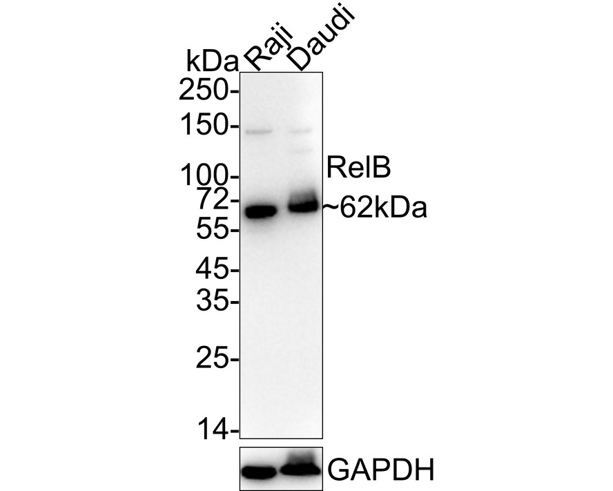
Western blot analysis of RelB on different lysates with Rabbit anti-RelB antibody at 1/1,000 dilution. Lane 1: Raji cell lysate, Lane 2: Daudi cell lysate, Lysates/proteins at 20 µg/Lane. Exposure time: 3 minutes; 4-20% SDS-PAGE gel. Proteins were transferred to a PVDF membrane and blocked with 5% NFDM/TBST for 1 hour at room temperature. The primary antibody at 1/1,000 dilution was used in 5% NFDM/TBST at 4℃ overnight. Goat Anti-Rabbit IgG - HRP Secondary Antibody at 1/50,000 dilution was used for 1 hour at room temperature.IHC
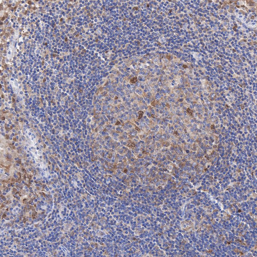
Immunohistochemical analysis of paraffin-embedded human tonsil tissue with Rabbit anti-RelB antibody at 1/50 dilution. The section was pre-treated using heat mediated antigen retrieval with sodium citrate buffer (pH 6.0) (high pressure) for 2 minutes. The tissues were blocked in 1% BSA for 20 minutes at room temperature, washed with ddH2O and PBS, and then probed with the primary antibody at 1/50 dilution for 1 hour at room temperature. The detection was performed using an HRP conjugated compact polymer system. DAB was used as the chromogen. Tissues were counterstained with hematoxylin and mounted with DPX.| Product Name | RelB Recombinant Rabbit Monoclonal Antibody |
|---|---|
| Antibody Type | Primary Antibodies |
| Product description | The NFκB transcription factor was originally identified as a protein complex consisting of a DNA binding subunit and an associated protein. The DNA binding subunit is functionally related to c-Rel p75 and Rel B p68. The p50 subunit was initially believed to be a functionally unique protein derived from the amino terminus of a precursor designated p105. A second protein designated p52 (previously referred to as p49) has been identified that can act as an alternative NFκB subunit. Rel B does not bind with high affinity to NFkB sites, but heterodimers between Rel B and p50 bind with an affinity comparable to that of p50 NFκB homodimers. However, Rel B/p50 heterodimers, in contrast to NFκB heterodimers, transactivates transcription of promotors containing κB binding sites. |
| Immunogen | Synthetic peptide within Human RelB aa 1-50 / 579. |
| Clonality | Monoclonal |
|---|---|
| Isotype | IgG |
| Host Species | Recombinant rabbit |
| Tested Applications | IHCWB |
| WB:1:1000 IHC:1:50-1:200 |
|
| Species Reactivity | Human |
| Concentration | 1mg/ml |
| Purification | Protein A |
| Alternative Names | I REL antibody I-Rel antibody IREL antibody Nuclear factor of kappa light polypeptide gene enhancer in B cells 3 antibody relB antibody RELB_HUMAN antibody Reticuloendotheliosis viral oncogene homolog B antibody Transcription factor Rel B antibody Transcription factor RelB antibody v rel avian reticuloendotheliosis viral oncogene homolog B antibody v rel reticuloendotheliosis viral oncogene homolog B antibody |
|---|---|
| Molecular Weight(MW) | 62 kDa |
| Cellular Localization | Nucleus, Cytoplasm, cytoskeleton, microtubule organizing center, centrosome. |
| SwissProt ID | Q01201 |
|---|
WB
Western blot analysis of RelB on different lysates with Rabbit anti-RelB antibody at 1/1,000 dilution. Lane 1: Raji cell lysate, Lane 2: Daudi cell lysate, Lysates/proteins at 20 µg/Lane. Exposure time: 3 minutes; 4-20% SDS-PAGE gel. Proteins were transferred to a PVDF membrane and blocked with 5% NFDM/TBST for 1 hour at room temperature. The primary antibody at 1/1,000 dilution was used in 5% NFDM/TBST at 4℃ overnight. Goat Anti-Rabbit IgG - HRP Secondary Antibody at 1/50,000 dilution was used for 1 hour at room temperature.
IHC
Immunohistochemical analysis of paraffin-embedded human tonsil tissue with Rabbit anti-RelB antibody at 1/50 dilution. The section was pre-treated using heat mediated antigen retrieval with sodium citrate buffer (pH 6.0) (high pressure) for 2 minutes. The tissues were blocked in 1% BSA for 20 minutes at room temperature, washed with ddH2O and PBS, and then probed with the primary antibody at 1/50 dilution for 1 hour at room temperature. The detection was performed using an HRP conjugated compact polymer system. DAB was used as the chromogen. Tissues were counterstained with hematoxylin and mounted with DPX.| Positive Control | A549, MCF-7, SW480, human spleen tissue, human breast carcinoma tissue. |
|---|---|
| Application Notes | WB:1:1000 IHC:1:50-1:200 |
| Form | Liquid |
|---|---|
| Storage Instructions | Store at +4℃ after thawing. Aliquot store at -20℃ or -80℃. Avoid repeated freeze / thaw cycles. |
| Storage Buffer | 1*TBS (pH7.4), 1%BSA, 40%Glycerol. Preservative: 0.05% Sodium Azide. |


