Application
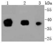
Fig1: Western blot analysis of Oct4 on different lysates using anti-Oct4 antibody at 1/1,000 dilution. Positive control: Lane 1: F9 Lane 2: NCCIT Lane 3: hESApplication
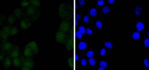
Fig2: ICC staining Oct4 in N2A cells (green). The nuclear counter stain is DAPI (blue). Cells were fixed in paraformaldehyde, permeabilised with 0.25% Triton X100/PBS.Application
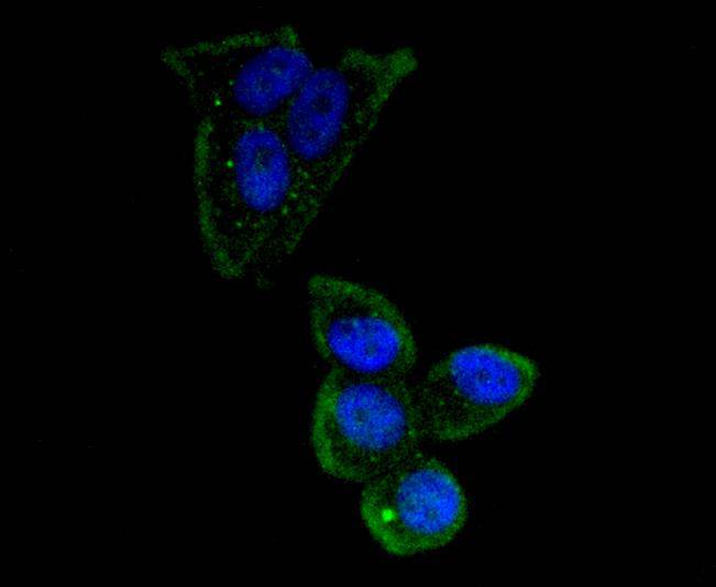
Fig3: ICC staining Oct4 in MCF-7 cells (green). The nuclear counter stain is DAPI (blue). Cells were fixed in paraformaldehyde, permeabilised with 0.25% Triton X100/PBS.Application
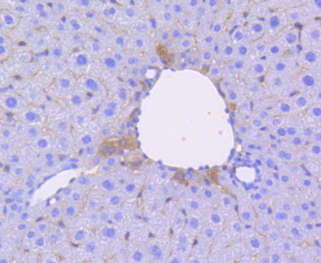
Fig4: Immunohistochemical analysis of paraffin-embedded mouse liver tissue using anti-Oct4 antibody. Counter stained with hematoxylin.| Product Name | Oct4 [SD0750] |
|---|---|
| Antibody Type | Primary Antibodies |
| Antigen Alias | Octamer binding transcription factor 4 antibody MGC22487 antibody Oct 3 antibody Oct 4 antibody Oct-3 antibody Oct-4 antibody OCT3 antibody Oct4 antibody Octamer binding protein 3 antibody Octamer binding protein 4 antibody Octamer binding transcription factor 3 antibody Octamer-binding protein 3 antibody Octamer-binding protein 4 antibody Octamer-binding transcription factor 3 antibody OTF 3 antibody OTF 4 antibody OTF-3 antibody OTF3 antibody OTF4 antibody PO5F1_HUMAN antibody POU class 5 homeobox 1 antibody POU domain class 5 transcription factor 1 antibody POU domain transcription factor OCT4 antibody POU domain, class 5, transcription factor 1 antibody POU-type homeodomain-containing DNA-binding protein antibody POU5F1 antibody |
| Product description | POU5F1 (POU domain, class 5, transcription factor 1), also known as octamer-binding transcription factor-3 (Oct-3, OTF3), octamer-binding transcription factor-4 (Oct-4, Otf-4) and Oct-3/4, modulates embryonic stem (ES) cell populations by influencing lineage commitment. Oct-3/4 sustains stem-cell self-renewal and differentiation pathways. Transcription factors containing the POU homeodomain regulate tissue-specific gene expression in lymphoid and pituitary differentiation and in early mammalian development. Oct-3/4 is capable of inducing rapid proliferation and tumorigenic properties of ES cells through activation of the UTF1 gene. In humans, two Oct-3/4 isoforms contribute to influencing the undifferentiated phenotype of ES cells. Oct-3/4 pseudogenes localizing to human chromosomes 10 and 8 are reported to be transcribed in certain cancer cell lines and tissues. |
| Immunogen | recombinant protein |
| Clonality | Monoclonal |
|---|---|
| Isotype | IgG |
| Host Species | Recombinant rabbit |
| Tested Applications | WBICC/IFIHCIPChIP |
| WB:1:1,000-1:5,000 ICC:1:50-1:200 IHC:1:50-1:200 |
|
| Species Reactivity | HumanMouse |
| Concentration | 1mg/ml |
| Alternative Names | Octamer binding transcription factor 4 antibody
MGC22487 antibody Oct 3 antibody Oct 4 antibody Oct-3 antibody Oct-4 antibody OCT3 antibody Oct4 antibody Octamer binding protein 3 antibody Octamer binding protein 4 antibody Octamer binding transcription factor 3 antibody Octamer-binding protein 3 antibody Octamer-binding protein 4 antibody Octamer-binding transcription factor 3 antibody OTF 3 antibody OTF 4 antibody OTF-3 antibody OTF3 antibody OTF4 antibody PO5F1_HUMAN antibody POU class 5 homeobox 1 antibody POU domain class 5 transcription factor 1 antibody POU domain transcription factor OCT4 antibody POU domain class 5 transcription factor 1 antibody POU-type homeodomain-containing DNA-binding protein antibody POU5F1 antibody |
|---|---|
| Molecular Weight(MW) | 39 kDa |
| Cellular Localization | Nucleus, Cytoplasm. |
Application
Fig1: Western blot analysis of Oct4 on different lysates using anti-Oct4 antibody at 1/1,000 dilution. Positive control: Lane 1: F9 Lane 2: NCCIT Lane 3: hES
Application
Fig2: ICC staining Oct4 in N2A cells (green). The nuclear counter stain is DAPI (blue). Cells were fixed in paraformaldehyde, permeabilised with 0.25% Triton X100/PBS.
Application
Fig3: ICC staining Oct4 in MCF-7 cells (green). The nuclear counter stain is DAPI (blue). Cells were fixed in paraformaldehyde, permeabilised with 0.25% Triton X100/PBS.
Application
Fig4: Immunohistochemical analysis of paraffin-embedded mouse liver tissue using anti-Oct4 antibody. Counter stained with hematoxylin.| Positive Control | F9, NCCIT, hES, N2A, MCF-7, mouse liver tissue. |
|---|---|
| Application Notes | WB:1:1,000-1:5,000 ICC:1:50-1:200 IHC:1:50-1:200 |
| Form | Liquid |
|---|---|
| Storage Instructions | Store at +4℃ after thawing. Aliquot store at -20℃ or -80℃. Avoid repeated freeze / thaw cycles. |
| Storage Buffer | 1*TBS (pH7.4), 1%BSA, 40%Glycerol. Preservative: 0.05% Sodium Azide. |


