WB
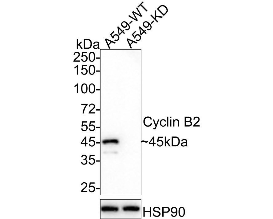
Western blot analysis of Cyclin B2 on different lysates with Rabbit anti-Cyclin B2 antibody at 1/1,000 dilution. Lane 1: A549-WT cell lysate, Lane 2: A549-KD Cyclin B2 cell lysate, Lysates/proteins at 10 µg/Lane. Exposure time: 3 minutes; 4-20% SDS-PAGE gel. Proteins were transferred to a PVDF membrane and blocked with 5% NFDM/TBST for 1 hour at room temperature. The primary antibody at 1/1,000 dilution was used in 5% NFDM/TBST at 4℃ overnight. Goat Anti-Rabbit IgG - HRP Secondary Antibody at 1/50,000 dilution was used for 1 hour at room temperature.WB
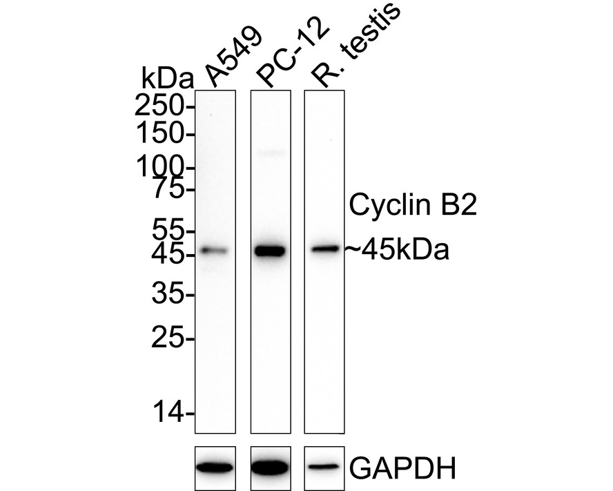
Western blot analysis of Cyclin B2 on different lysates with Rabbit anti-Cyclin B2 antibody at 1/1,000 dilution. Lane 1: A549 cell lysate (20 µg/Lane), Lane 2: PC-12 cell lysate (20 µg/Lane), Lane 3: Rat testis tissue lysate (40 µg/Lane), Exposure time: 3 minutes; 4-20% SDS-PAGE gel. Proteins were transferred to a PVDF membrane and blocked with 5% NFDM/TBST for 1 hour at room temperature. The primary antibody at 1/1,000 dilution was used in 5% NFDM/TBST at 4℃ overnight. Goat Anti-Rabbit IgG - HRP Secondary Antibody at 1/50,000 dilution was used for 1 hour at room temperature.ICC/IF
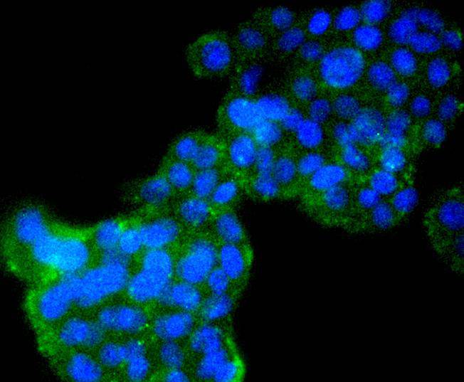
ICC staining Cyclin B2 in PC-12 cells (green). The nuclear counter stain is DAPI (blue). Cells were fixed in paraformaldehyde, permeabilised with 0.25% Triton X100/PBS.| Product Name | Cyclin B2 Recombinant Rabbit Monoclonal Antibody |
|---|---|
| Antibody Type | Primary Antibodies |
| Product description | In eukaryotic cells, mitosis is initiated following the activation of a protein kinase known variously as maturation-promoting factor, M-phase specific histone kinase or M-phase kinase. This protein kinase is composed of a catalytic subunit (Cdc2), a regulatory subunit (cyclin B) and a low molecular weight subunit (p13-Suc 1). The Cdc/cyclin enzyme is subject to multiple levels of control of which the regulation of the catalytic subunit by tyrosine phosphorylation is the best understood. Tyrosine phosphorylation inhibits the Cdc2/cyclin B enzyme and tyrosine dephosphorylation, occurring at the onset of mitosis, directly activates the pre-MPF complex . Evidence has established that B-type cyclins not only act on M-phase regulatory subunits of the Cdc2 protein kinase, but also activate the Cdc25A and Cdc25B endogenous tyrosine phosphatase, of which Cdc2 is the physiological substrate. The two B-type cyclins, cyclin B1 and cyclin B2, have been shown to have distinct tissue distributions. |
| Immunogen | Recombinant protein within Human Cyclin B2 aa 1-102 / 398. |
| Clonality | Monoclonal |
|---|---|
| Isotype | IgG |
| Host Species | Recombinant rabbit |
| Tested Applications | ICC/IFWB |
| WB:1:1000 ICC/IF:1:50-1:200 |
|
| Species Reactivity | HumanRat |
| Concentration | 1mg/ml |
| Purification | Protein A |
| Alternative Names | ccnb2 antibody CCNB2_HUMAN antibody CycB2 antibody Cyclin B2 antibody G2 mitotic specific cyclin B2 antibody G2/mitotic specific cyclin B2 antibody G2/mitotic-specific cyclin-B2 antibody HsT17299 antibody MGC108931 antibody MGC140694 antibody |
|---|---|
| Molecular Weight(MW) | 45 kDa |
| Cellular Localization | Centrosome, microtubule cytoskeleton, cytosol, nucleus, cyclin-dependent protein kinase holoenzyme complex, cytoplasm, host cell nucleus, membrane. |
WB
Western blot analysis of Cyclin B2 on different lysates with Rabbit anti-Cyclin B2 antibody at 1/1,000 dilution. Lane 1: A549-WT cell lysate, Lane 2: A549-KD Cyclin B2 cell lysate, Lysates/proteins at 10 µg/Lane. Exposure time: 3 minutes; 4-20% SDS-PAGE gel. Proteins were transferred to a PVDF membrane and blocked with 5% NFDM/TBST for 1 hour at room temperature. The primary antibody at 1/1,000 dilution was used in 5% NFDM/TBST at 4℃ overnight. Goat Anti-Rabbit IgG - HRP Secondary Antibody at 1/50,000 dilution was used for 1 hour at room temperature.
WB
Western blot analysis of Cyclin B2 on different lysates with Rabbit anti-Cyclin B2 antibody at 1/1,000 dilution. Lane 1: A549 cell lysate (20 µg/Lane), Lane 2: PC-12 cell lysate (20 µg/Lane), Lane 3: Rat testis tissue lysate (40 µg/Lane), Exposure time: 3 minutes; 4-20% SDS-PAGE gel. Proteins were transferred to a PVDF membrane and blocked with 5% NFDM/TBST for 1 hour at room temperature. The primary antibody at 1/1,000 dilution was used in 5% NFDM/TBST at 4℃ overnight. Goat Anti-Rabbit IgG - HRP Secondary Antibody at 1/50,000 dilution was used for 1 hour at room temperature.
ICC/IF
ICC staining Cyclin B2 in PC-12 cells (green). The nuclear counter stain is DAPI (blue). Cells were fixed in paraformaldehyde, permeabilised with 0.25% Triton X100/PBS.| Application Notes | WB:1:1000 ICC/IF:1:50-1:200 |
|---|
| Form | Liquid |
|---|---|
| Storage Instructions | Store at +4℃ after thawing. Aliquot store at -20℃ or -80℃. Avoid repeated freeze / thaw cycles. |
| Storage Buffer | 1*TBS (pH7.4), 1%BSA, 40%Glycerol. Preservative: 0.05% Sodium Azide. |


