WB
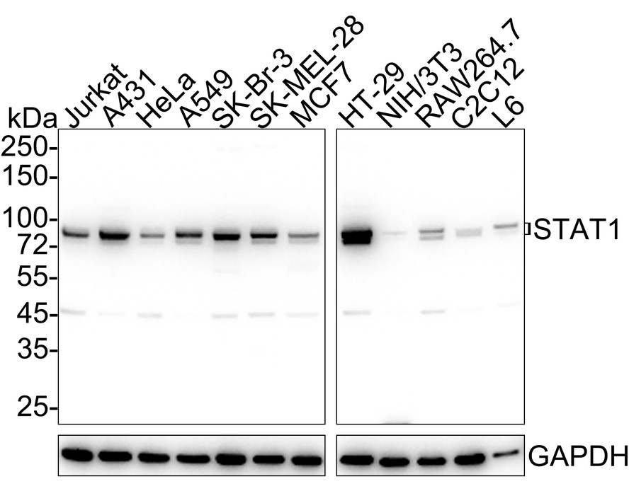
Western blot analysis of STAT1 on different lysates with Rabbit anti-STAT1 antibody at 1/1,000 dilution. Lane 1: Jurkat cell lysate (15 µg/Lane), Lane 2: A431 cell lysate (15 µg/Lane), Lane 3: HeLa cell lysate (15 µg/Lane), Lane 4: A549 cell lysate (15 µg/Lane), Lane 5: SK-Br-3 cell lysate (15 µg/Lane), Lane 6: SK-MEL-28 cell lysate (15 µg/Lane), Lane 7: MCF7 cell lysate (15 µg/Lane), Lane 8: HT-29 cell lysate (15 µg/Lane), Lane 9: NIH/3T3 cell lysate (15 µg/Lane), Lane 10: RAW264.7 cell lysate (15 µg/Lane), Lane 11: C2C12 cell lysate (15 µg/Lane), Lane 12: L6 cell lysate (15 µg/Lane), Exposure time: 10 seconds; 4-20% SDS-PAGE gel. Proteins were transferred to a PVDF membrane and blocked with 5% NFDM/TBST for 1 hour at room temperature. The primary antibody at 1/1,000 dilution was used in 5% NFDM/TBST at 4℃ overnight. Goat Anti-Rabbit IgG - HRP Secondary Antibody at 1/50,000 dilution was used for 1 hour at room temperature.IHC
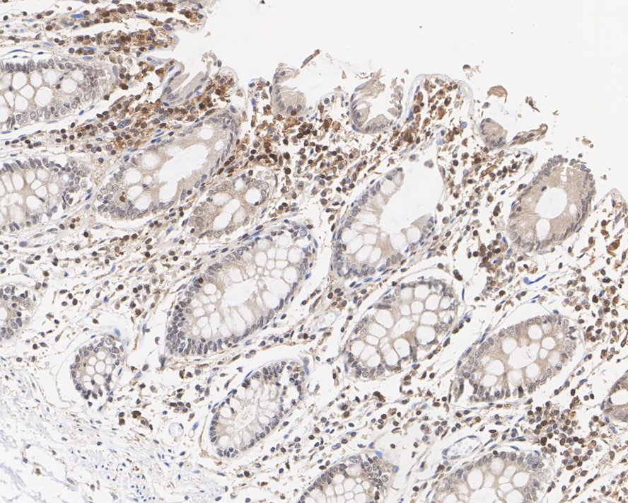
Immunohistochemical analysis of paraffin-embedded human colon tissue with Rabbit anti-STAT1 antibody at 1/500 dilution. The section was pre-treated using heat mediated antigen retrieval with sodium citrate buffer (pH 6.0) for 2 minutes. The tissues were blocked in 1% BSA for 20 minutes at room temperature, washed with ddH2O and PBS, and then probed with the primary antibody at 1/500 dilution for 1 hour at room temperature. The detection was performed using an HRP conjugated compact polymer system. DAB was used as the chromogen. Tissues were counterstained with hematoxylin and mounted with DPX.IHC
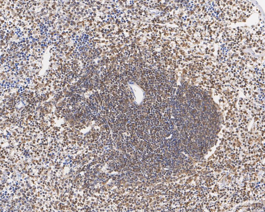
Immunohistochemical analysis of paraffin-embedded mouse spleen tissue with Rabbit anti-STAT1 antibody at 1/2,000 dilution. The section was pre-treated using heat mediated antigen retrieval with sodium citrate buffer (pH 6.0) for 2 minutes. The tissues were blocked in 1% BSA for 20 minutes at room temperature, washed with ddH2O and PBS, and then probed with the primary antibody at 1/2,000 dilution for 1 hour at room temperature. The detection was performed using an HRP conjugated compact polymer system. DAB was used as the chromogen. Tissues were counterstained with hematoxylin and mounted with DPX.IHC
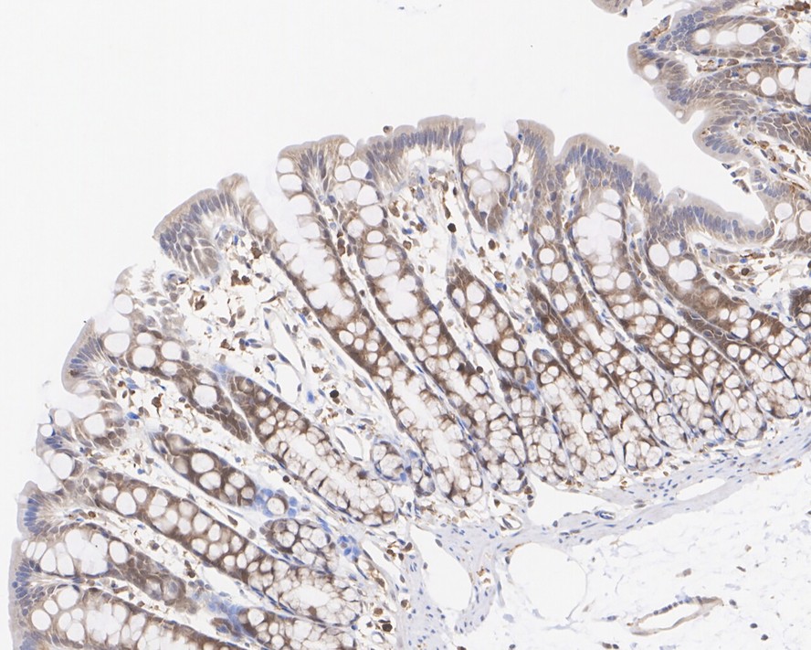
Immunohistochemical analysis of paraffin-embedded rat colon tissue with Rabbit anti-STAT1 antibody at 1/2,000 dilution. The section was pre-treated using heat mediated antigen retrieval with sodium citrate buffer (pH 6.0) for 2 minutes. The tissues were blocked in 1% BSA for 20 minutes at room temperature, washed with ddH2O and PBS, and then probed with the primary antibody at 1/2,000 dilution for 1 hour at room temperature. The detection was performed using an HRP conjugated compact polymer system. DAB was used as the chromogen. Tissues were counterstained with hematoxylin and mounted with DPX.ICC/IF
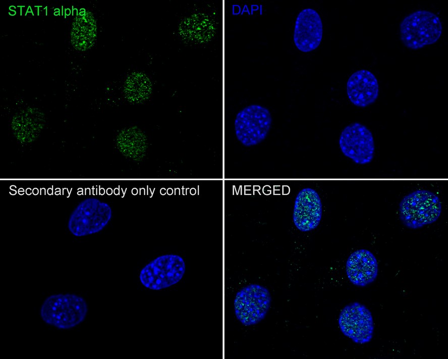
Immunocytochemistry analysis of NIH/3T3 cells labeling STAT1 with Rabbit anti-STAT1 antibody at 1/100 dilution. Cells were fixed in 4% paraformaldehyde for 20 minutes at room temperature, permeabilized with 0.1% Triton X-100 in PBS for 5 minutes at room temperature, then blocked with 1% BSA in 10% negative goat serum for 1 hour at room temperature. Cells were then incubated with Rabbit anti-STAT1 antibody at 1/100 dilution in 1% BSA in PBST overnight at 4 ℃. Goat Anti-Rabbit IgG H&L (488) was used as the secondary antibody at 1/1,000 dilution. PBS instead of the primary antibody was used as the secondary antibody only control. Nuclear DNA was labelled in blue with DAPI.IF-P
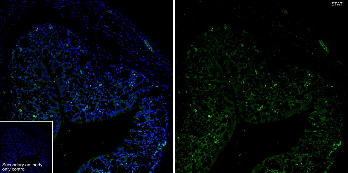
Immunofluorescence analysis of paraffin-embedded mouse colon tissue labeling STAT1 with Rabbit anti-STAT1 antibody at 1/200 dilution. The section was pre-treated using heat mediated antigen retrieval with sodium citrate buffer (pH 6.0) for 2 minutes. The tissues were blocked in 10% negative goat serum for 1 hour at room temperature, washed with PBS, and then probed with the primary antibody (green) at 1/200 dilution overnight at 4 ℃, washed with PBS. Goat Anti-Rabbit IgG H&L (488) was used as the secondary antibody at 1/1,000 dilution. Nuclei were counterstained with DAPI (blue).FC
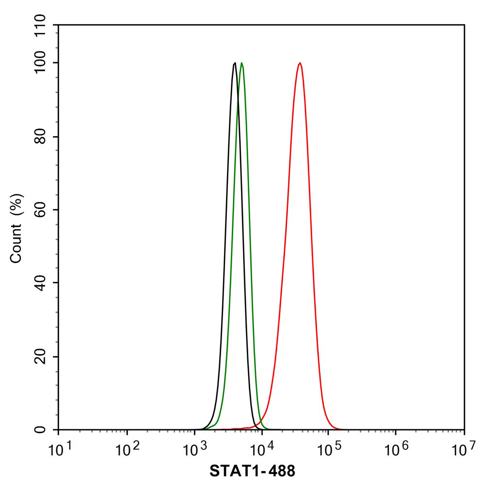
Flow cytometric analysis of NIH/3T3 cells labeling STAT1. Cells were fixed and permeabilized. Then stained with the primary antibody (1/1,000) (red) compared with Rabbit IgG Isotype Control (green). After incubation of the primary antibody at +4℃ for an hour, the cells were stained with a 488 conjugate-Goat anti-Rabbit IgG Secondary antibody at 1/1,000 dilution for 30 minutes at +4℃. Unlabelled sample was used as a control (cells without incubation with primary antibody; black).IP
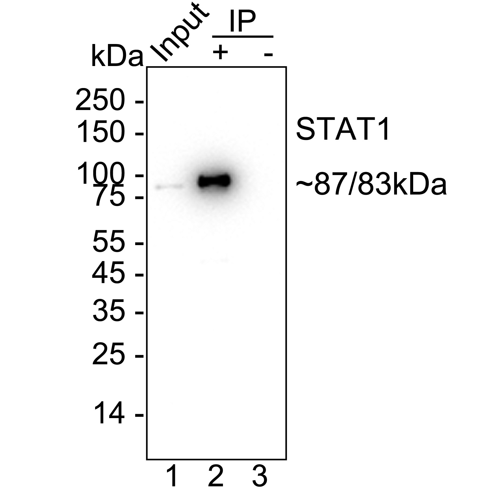
STAT1 was immunoprecipitated from 0.2 mg HeLa cell lysate with Rabbit anti-STAT1 antibody at 2 µg/25 µl agarose. Western blot was performed from the immunoprecipitate using Rabbit anti-STAT1 antibody at 1/1,000 dilution. Anti-Rabbit IgG for IP Nano-secondary antibody at 1/5,000 dilution was used for 1 hour at room temperature. Lane 1: HeLa cell lysate (input), Lane 2: Rabbit anti-STAT1 antibody IP in HeLa cell lysate, Lane 3: Rabbit IgG instead of Rabbit anti-STAT1 antibody in HeLa cell lysate, Blocking/Dilution buffer: 5% NFDM/TBST, Exposure time: 12 seconds.| Product Name | STAT1 Recombinant Rabbit Monoclonal Antibody |
|---|---|
| Antibody Type | Primary Antibodies |
| Product description | Membrane receptor signaling by various ligands, including interferons and growth hormones such as EGF, induces activation of JAK kinases which then leads to tyrosine phosphorylation of the various Stat transcription factors. Stat1 and Stat2 are induced by IFN-α and form a heterodimer which is part of the ISGF3 transcription factor complex. Although early reports indicate Stat3 activation by EGF and IL-6, it has been shown that Stat3β appears to be activated by both while Stat3α is activated by EGF, but not by IL-6. Highest expression of Stat4 is seen in testis and myeloid cells. IL-12 has been identified as an activator of Stat4. Stat5 has been shown to be activated by Prolactin and by IL-3. Stat6 is involved in IL-4 activated signaling pathways. |
| Immunogen | Synthetic peptide within Human STAT1 alpha aa 1-50 / 750. |
| Clonality | Monoclonal |
|---|---|
| Isotype | IgG |
| Host Species | Recombinant rabbit |
| Tested Applications | FCICC/IFIF-PIHCIPWB |
| WB:1:1000-1:5000 IHC:1:500-1:2000 ICC/IF:1:50-1:200 IF-P:1:200 FC:1:1000 IP:1-2μg/sample. |
|
| Species Reactivity | HumanMouseRat |
| Concentration | 1mg/ml |
| Purification | Protein A |
| Alternative Names | CANDF7 antibody
DKFZp686B04100 antibody
ISGF 3 antibody
ISGF3 antibody
OTTHUMP00000163552 antibody
OTTHUMP00000165046 antibody
OTTHUMP00000165047 antibody
OTTHUMP00000205845 antibody
Signal transducer and activator of transcription 1 antibody
Signal transducer and activator of transcription 1 91kDa antibody Signal transducer and activator of transcription 1-alpha/beta antibody Stat1 antibody STAT1_HUMAN antibody STAT91 antibody Transcription factor ISGF-3 components p91/p84 antibody |
|---|---|
| Molecular Weight(MW) | 87/83kDa |
| Cellular Localization | Cytoplasm, Nucleus. |
| SwissProt ID | P42224 |
|---|
WB
Western blot analysis of STAT1 on different lysates with Rabbit anti-STAT1 antibody at 1/1,000 dilution. Lane 1: Jurkat cell lysate (15 µg/Lane), Lane 2: A431 cell lysate (15 µg/Lane), Lane 3: HeLa cell lysate (15 µg/Lane), Lane 4: A549 cell lysate (15 µg/Lane), Lane 5: SK-Br-3 cell lysate (15 µg/Lane), Lane 6: SK-MEL-28 cell lysate (15 µg/Lane), Lane 7: MCF7 cell lysate (15 µg/Lane), Lane 8: HT-29 cell lysate (15 µg/Lane), Lane 9: NIH/3T3 cell lysate (15 µg/Lane), Lane 10: RAW264.7 cell lysate (15 µg/Lane), Lane 11: C2C12 cell lysate (15 µg/Lane), Lane 12: L6 cell lysate (15 µg/Lane), Exposure time: 10 seconds; 4-20% SDS-PAGE gel. Proteins were transferred to a PVDF membrane and blocked with 5% NFDM/TBST for 1 hour at room temperature. The primary antibody at 1/1,000 dilution was used in 5% NFDM/TBST at 4℃ overnight. Goat Anti-Rabbit IgG - HRP Secondary Antibody at 1/50,000 dilution was used for 1 hour at room temperature.
IHC
Immunohistochemical analysis of paraffin-embedded human colon tissue with Rabbit anti-STAT1 antibody at 1/500 dilution. The section was pre-treated using heat mediated antigen retrieval with sodium citrate buffer (pH 6.0) for 2 minutes. The tissues were blocked in 1% BSA for 20 minutes at room temperature, washed with ddH2O and PBS, and then probed with the primary antibody at 1/500 dilution for 1 hour at room temperature. The detection was performed using an HRP conjugated compact polymer system. DAB was used as the chromogen. Tissues were counterstained with hematoxylin and mounted with DPX.
IHC
Immunohistochemical analysis of paraffin-embedded mouse spleen tissue with Rabbit anti-STAT1 antibody at 1/2,000 dilution. The section was pre-treated using heat mediated antigen retrieval with sodium citrate buffer (pH 6.0) for 2 minutes. The tissues were blocked in 1% BSA for 20 minutes at room temperature, washed with ddH2O and PBS, and then probed with the primary antibody at 1/2,000 dilution for 1 hour at room temperature. The detection was performed using an HRP conjugated compact polymer system. DAB was used as the chromogen. Tissues were counterstained with hematoxylin and mounted with DPX.
IHC
Immunohistochemical analysis of paraffin-embedded rat colon tissue with Rabbit anti-STAT1 antibody at 1/2,000 dilution. The section was pre-treated using heat mediated antigen retrieval with sodium citrate buffer (pH 6.0) for 2 minutes. The tissues were blocked in 1% BSA for 20 minutes at room temperature, washed with ddH2O and PBS, and then probed with the primary antibody at 1/2,000 dilution for 1 hour at room temperature. The detection was performed using an HRP conjugated compact polymer system. DAB was used as the chromogen. Tissues were counterstained with hematoxylin and mounted with DPX.
ICC/IF
Immunocytochemistry analysis of NIH/3T3 cells labeling STAT1 with Rabbit anti-STAT1 antibody at 1/100 dilution. Cells were fixed in 4% paraformaldehyde for 20 minutes at room temperature, permeabilized with 0.1% Triton X-100 in PBS for 5 minutes at room temperature, then blocked with 1% BSA in 10% negative goat serum for 1 hour at room temperature. Cells were then incubated with Rabbit anti-STAT1 antibody at 1/100 dilution in 1% BSA in PBST overnight at 4 ℃. Goat Anti-Rabbit IgG H&L (488) was used as the secondary antibody at 1/1,000 dilution. PBS instead of the primary antibody was used as the secondary antibody only control. Nuclear DNA was labelled in blue with DAPI.
IF-P
Immunofluorescence analysis of paraffin-embedded mouse colon tissue labeling STAT1 with Rabbit anti-STAT1 antibody at 1/200 dilution. The section was pre-treated using heat mediated antigen retrieval with sodium citrate buffer (pH 6.0) for 2 minutes. The tissues were blocked in 10% negative goat serum for 1 hour at room temperature, washed with PBS, and then probed with the primary antibody (green) at 1/200 dilution overnight at 4 ℃, washed with PBS. Goat Anti-Rabbit IgG H&L (488) was used as the secondary antibody at 1/1,000 dilution. Nuclei were counterstained with DAPI (blue).
FC
Flow cytometric analysis of NIH/3T3 cells labeling STAT1. Cells were fixed and permeabilized. Then stained with the primary antibody (1/1,000) (red) compared with Rabbit IgG Isotype Control (green). After incubation of the primary antibody at +4℃ for an hour, the cells were stained with a 488 conjugate-Goat anti-Rabbit IgG Secondary antibody at 1/1,000 dilution for 30 minutes at +4℃. Unlabelled sample was used as a control (cells without incubation with primary antibody; black).
IP
STAT1 was immunoprecipitated from 0.2 mg HeLa cell lysate with Rabbit anti-STAT1 antibody at 2 µg/25 µl agarose. Western blot was performed from the immunoprecipitate using Rabbit anti-STAT1 antibody at 1/1,000 dilution. Anti-Rabbit IgG for IP Nano-secondary antibody at 1/5,000 dilution was used for 1 hour at room temperature. Lane 1: HeLa cell lysate (input), Lane 2: Rabbit anti-STAT1 antibody IP in HeLa cell lysate, Lane 3: Rabbit IgG instead of Rabbit anti-STAT1 antibody in HeLa cell lysate, Blocking/Dilution buffer: 5% NFDM/TBST, Exposure time: 12 seconds.| Application Notes | WB:1:1000-1:5000 IHC:1:500-1:2000 ICC/IF:1:50-1:200 IF-P:1:200 FC:1:1000 IP:1-2μg/sample. |
|---|
| Form | Liquid |
|---|---|
| Storage Instructions | Store at +4℃ after thawing. Aliquot store at -20℃ or -80℃. Avoid repeated freeze / thaw cycles. |
| Storage Buffer | 1*TBS (pH7.4), 1%BSA, 40%Glycerol. Preservative: 0.05% Sodium Azide. |


