Application
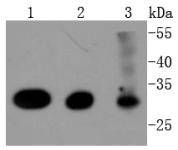
Fig1: Western blot analysis of Cdk4 on different lysates using anti-Cdk4 antibody at 1/1,000 dilution. Positive control: Lane 1: Hela Lane 2: MCF-7 Lane 3: K562Application
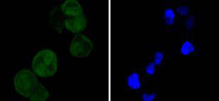
Fig2: ICC staining Cdk4 in Hela cells (green). The nuclear counter stain is DAPI (blue). Cells were fixed in paraformaldehyde, permeabilised with 0.25% Triton X100/PBS.Application
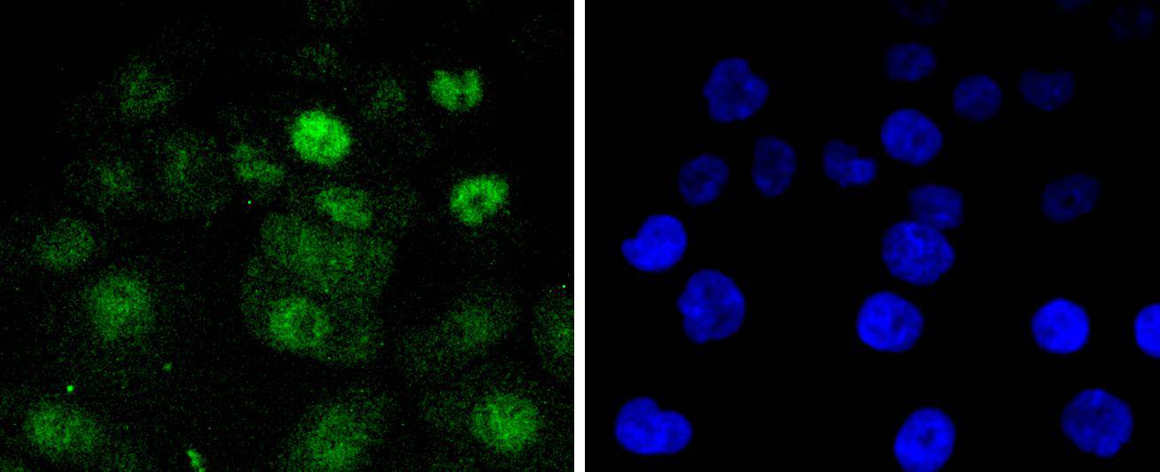
Fig3: ICC staining Cdk4 in AGS cells (green). The nuclear counter stain is DAPI (blue). Cells were fixed in paraformaldehyde, permeabilised with 0.25% Triton X100/PBS.Application
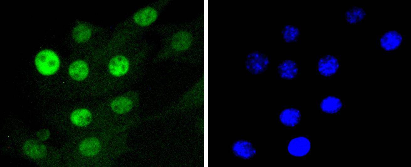
Fig4: ICC staining Cdk4 in NIH/3T3 cells (green). The nuclear counter stain is DAPI (blue). Cells were fixed in paraformaldehyde, permeabilised with 0.25% Triton X100/PBS.| Product Name | Cdk4 [SD205-1] |
|---|---|
| Antibody Type | Primary Antibodies |
| Antigen Alias | Cdk 4 antibody cdk4 antibody CDK4 protein antibody CDK4_HUMAN antibody Cell division kinase 4 antibody Cell division protein kinase 4 antibody CMM 3 antibody CMM3 antibody Crk3 antibody Cyclin dependent kinase 4 antibody Cyclin-dependent kinase 4 antibody Melanoma cutaneous malignant 3 antibody MGC14458 antibody p34 cdk4 antibody PSK J3 antibody PSK-J3 antibody |
| Product description | Cell cycle progression is controlled in part by a family of cyclin proteins and cyclin dependent kinases (Cdks). Cdk proteins work in concert with the cyclins to phosphorylate key substrates involved in each phase of cell cycle progression. Another family of proteins, Cdk inhibitors, also plays a role in regulating the cell cycle by binding to cyclin-Cdk complexes and modulating their activity. Several Cdk proteins have been identified, including Cdk2-Cdk8, PCTAIRE-1-PCTAIRE-3, PITALRE and PITSLRE. Cdk4, in complex with D-type cyclins, is thought to regulate cell growth during the G1 phase of the cell cycle. This association with a D-type cyclin upregulates Cdk4 activity, whereas binding to the Cdk inhibitor p16 downregulates Cdk4 activity. Activation of the Cdk4-cyclin complexes requires phosphorylation on a single threonyl residue of Cdk4, catalyzed by a Cdk-activating protein (CAK). |
| Immunogen | recombinant protein |
| Clonality | Monoclonal |
|---|---|
| Isotype | IgG |
| Host Species | Recombinant rabbit |
| Tested Applications | WBICC/IFIP |
| WB:1:1,000-1:2,000 ICC:1:50-1:200 |
|
| Species Reactivity | HumanMouseRat |
| Concentration | 1mg/ml |
| Alternative Names | Cdk 4 antibody cdk4 antibody CDK4 protein antibody CDK4_HUMAN antibody Cell division kinase 4 antibody Cell division protein kinase 4 antibody CMM 3 antibody CMM3 antibody Crk3 antibody Cyclin dependent kinase 4 antibody Cyclin-dependent kinase 4 antibody Melanoma cutaneous malignant 3 antibody MGC14458 antibody p34 cdk4 antibody PSK J3 antibody PSK-J3 antibody |
|---|---|
| Molecular Weight(MW) | 34 kDa |
| Cellular Localization | Cytoplasm, Nucleus, Membrane. |
Application
Fig1: Western blot analysis of Cdk4 on different lysates using anti-Cdk4 antibody at 1/1,000 dilution. Positive control: Lane 1: Hela Lane 2: MCF-7 Lane 3: K562
Application
Fig2: ICC staining Cdk4 in Hela cells (green). The nuclear counter stain is DAPI (blue). Cells were fixed in paraformaldehyde, permeabilised with 0.25% Triton X100/PBS.
Application
Fig3: ICC staining Cdk4 in AGS cells (green). The nuclear counter stain is DAPI (blue). Cells were fixed in paraformaldehyde, permeabilised with 0.25% Triton X100/PBS.
Application
Fig4: ICC staining Cdk4 in NIH/3T3 cells (green). The nuclear counter stain is DAPI (blue). Cells were fixed in paraformaldehyde, permeabilised with 0.25% Triton X100/PBS.| Positive Control | K562, MCF-7, Hela, AGS, NIH/3T3. |
|---|---|
| Application Notes | WB:1:1,000-1:2,000 ICC:1:50-1:200 |
| Form | Liquid |
|---|---|
| Storage Instructions | Store at +4℃ after thawing. Aliquot store at -20℃ or -80℃. Avoid repeated freeze / thaw cycles. |
| Storage Buffer | 1*TBS (pH7.4), 1%BSA, 40%Glycerol. Preservative: 0.05% Sodium Azide. |


