Application
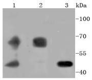
Fig1: Western blot analysis of PDPK1 on different lysates using anti-PDPK1 antibody at 1/1,000 dilution. Positive control: Lane 1: 293T Lane 2: PC-12 Lane 3: HL-60Application
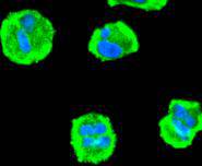
Fig2: ICC staining PDPK1 in Hela cells (green). The nuclear counter stain is DAPI (blue). Cells were fixed in paraformaldehyde, permeabilised with 0.25% Triton X100/PBS.Application
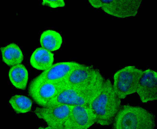
Fig3: ICC staining PDPK1 in MCF-7 cells (green). The nuclear counter stain is DAPI (blue). Cells were fixed in paraformaldehyde, permeabilised with 0.25% Triton X100/PBS.Application
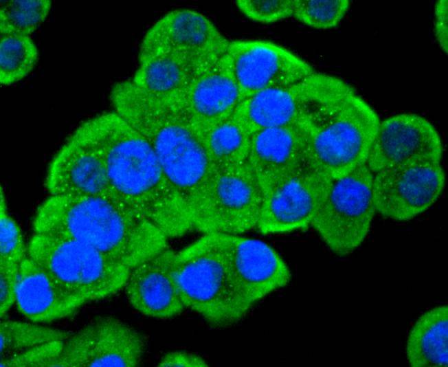
Fig4: ICC staining PDPK1 in SW480 cells (green). The nuclear counter stain is DAPI (blue). Cells were fixed in paraformaldehyde, permeabilised with 0.25% Triton X100/PBS.Application
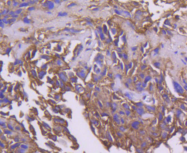
Fig5: Immunohistochemical analysis of paraffin-embedded human breast carcinoma tissue using anti-PDPK1 antibody. Counter stained with hematoxylin.Application
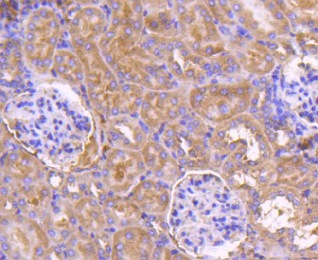
Fig6: Immunohistochemical analysis of paraffin-embedded mouse kidney tissue using anti-PDPK1 antibody. Counter stained with hematoxylin.Application
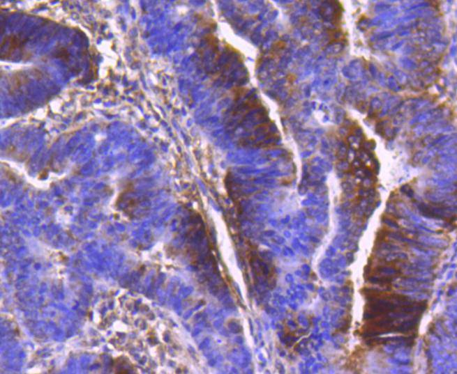
Fig7: Immunohistochemical analysis of paraffin-embedded human colon cancer tissue using anti-PDPK1 antibody. Counter stained with hematoxylin.Application
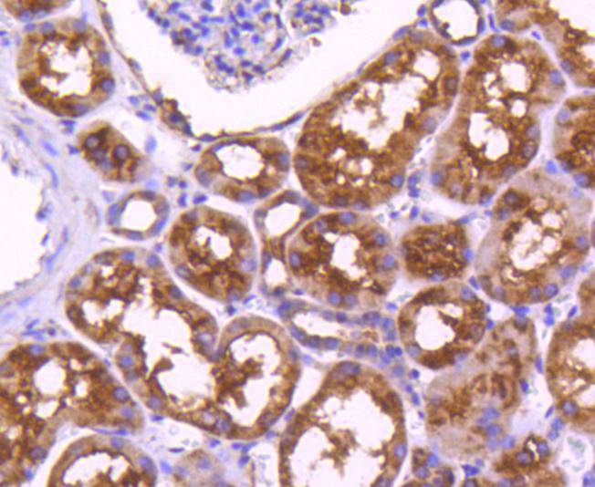
Fig8: Immunohistochemical analysis of paraffin-embedded human kidney tissue using anti-PDPK1 antibody. Counter stained with hematoxylin.| Product Name | PDPK1 [SD204-9] |
|---|---|
| Antibody Type | Primary Antibodies |
| Antigen Alias | 3 phosphoinositide dependent protein kinase 1 antibody 3-phosphoinositide-dependent protein kinase 1 antibody hPDK 1 antibody hPDK1 antibody MGC20087 antibody MGC35290 antibody OTTHUMP00000159109 antibody OTTHUMP00000159110 antibody OTTHUMP00000174525 antibody PDK1 antibody Pdpk1 antibody PDPK1_HUMAN antibody PDPK2 antibody PDPK2P antibody PkB kinase antibody PkB kinase like gene 1 antibody PkB like 1 antibody PRO0461 antibody Protein kinase antibody |
| Product description | PDPK1 (3-phosphoinositide dependent protein kinase 1), also known as PDK1, PDPK2, PDPK2P or PRO0461, is 556 amino acid ubiquitously expressed protein that localizes to the cell membrane, cytoplasm and nucleus. Acting as a master kinase, PDPK1 phosphorylates and activates a subgroup of the AGC family of protein kinases. PDPK1 is involved in mediating signal transduction for controlling proliferation, survival, and growth of developing pancreatic beta cells, regulating Ca2+ uptake and Ca2+-activated K+ channels of mast cells, regulation of chemotaxis and motility of vascular endothelial cells, cardiac homeostasis, and thymocyte development. Belonging to the protein kinase superfamily, PDPK1 contains a PH domain, which play an essential role in homodimerization, localization and nuclear import of PDPK1, and a protein kinase domain. PDPK1 exists as five alternatively spliced isoforms and is encoded by a gene located on human chromosome 16p13.3. |
| Immunogen | recombinant protein |
| Clonality | Monoclonal |
|---|---|
| Isotype | IgG |
| Host Species | Recombinant rabbit |
| Tested Applications | WBICC/IFIHC |
| WB:1:1,000-1:2,000 ICC:1:100-1:500 IHC:1:50-1:200 |
|
| Species Reactivity | HumanMouseRat |
| Concentration | 1mg/ml |
| Alternative Names | 3 phosphoinositide dependent protein kinase 1 antibody 3-phosphoinositide-dependent protein kinase 1 antibody hPDK 1 antibody hPDK1 antibody MGC20087 antibody MGC35290 antibody OTTHUMP00000159109 antibody OTTHUMP00000159110 antibody OTTHUMP00000174525 antibody PDK1 antibody Pdpk1 antibody PDPK1_HUMAN antibody PDPK2 antibody PDPK2P antibody PkB kinase antibody PkB kinase like gene 1 antibody PkB like 1 antibody PRO0461 antibody Protein kinase antibody |
|---|---|
| Molecular Weight(MW) | 63/48 kDa |
| Cellular Localization | Cytoplasm, Nucleus, Cell membrane, Cell junction. |
Application
Fig1: Western blot analysis of PDPK1 on different lysates using anti-PDPK1 antibody at 1/1,000 dilution. Positive control: Lane 1: 293T Lane 2: PC-12 Lane 3: HL-60
Application
Fig2: ICC staining PDPK1 in Hela cells (green). The nuclear counter stain is DAPI (blue). Cells were fixed in paraformaldehyde, permeabilised with 0.25% Triton X100/PBS.
Application
Fig3: ICC staining PDPK1 in MCF-7 cells (green). The nuclear counter stain is DAPI (blue). Cells were fixed in paraformaldehyde, permeabilised with 0.25% Triton X100/PBS.
Application
Fig4: ICC staining PDPK1 in SW480 cells (green). The nuclear counter stain is DAPI (blue). Cells were fixed in paraformaldehyde, permeabilised with 0.25% Triton X100/PBS.
Application
Fig5: Immunohistochemical analysis of paraffin-embedded human breast carcinoma tissue using anti-PDPK1 antibody. Counter stained with hematoxylin.
Application
Fig6: Immunohistochemical analysis of paraffin-embedded mouse kidney tissue using anti-PDPK1 antibody. Counter stained with hematoxylin.
Application
Fig7: Immunohistochemical analysis of paraffin-embedded human colon cancer tissue using anti-PDPK1 antibody. Counter stained with hematoxylin.
Application
Fig8: Immunohistochemical analysis of paraffin-embedded human kidney tissue using anti-PDPK1 antibody. Counter stained with hematoxylin.| Positive Control | 293T, PC-12, HL-60, Hela, MCF-7, SW480, human colon cancer tissue, human breast carcinoma tissue, human kidney tissue, mouse kidney tissue. |
|---|---|
| Application Notes | WB:1:1,000-1:2,000 ICC:1:100-1:500 IHC:1:50-1:200 |
| Form | Liquid |
|---|---|
| Storage Instructions | Store at +4℃ after thawing. Aliquot store at -20℃ or -80℃. Avoid repeated freeze / thaw cycles. |
| Storage Buffer | 1*TBS (pH7.4), 1%BSA, 40%Glycerol. Preservative: 0.05% Sodium Azide. |


