Application
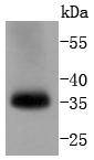
Fig1: Western blot analysis of pro Caspase 7 on human lung lysates using anti-pro Caspase 7 antibody at 1/1,000 dilution.Application
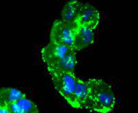
Fig2: ICC staining pro Caspase 7 in Hela cells (green). The nuclear counter stain is DAPI (blue). Cells were fixed in paraformaldehyde, permeabilised with 0.25% Triton X100/PBS.Application
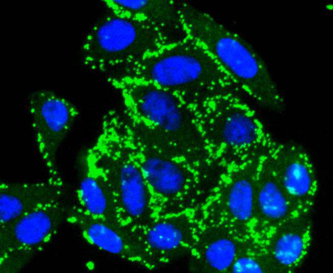
Fig3: ICC staining pro Caspase 7 in HepG2 cells (green). The nuclear counter stain is DAPI (blue). Cells were fixed in paraformaldehyde, permeabilised with 0.25% Triton X100/PBS.Application
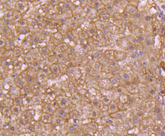
Fig4: Immunohistochemical analysis of paraffin-embedded human liver tissue using anti-pro Caspase 7 antibody. Counter stained with hematoxylin.Application
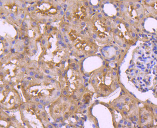
Fig5: Immunohistochemical analysis of paraffin-embedded human kidney tissue using anti-pro Caspase 7 antibody. Counter stained with hematoxylin.| Product Name | pro Caspase 7 [SD20-57] |
|---|---|
| Antibody Type | Primary Antibodies |
| Antigen Alias | apoptosis-related cysteine peptidase antibody Apoptotic protease Mch-3 antibody CASP-7 antibody CASP7 antibody CASP7_HUMAN antibody Caspase-7 subunit p11 antibody CMH-1 antibody ICE-LAP3 antibody ICE-like apoptotic protease 3 antibody |
| Product description | A unique family of Cysteine proteases has been described that differs in sequence, structure and substrate specificity from any previously described protease family. This family, Ced-3/caspase-1, is comprised of caspase-1, caspase-2, caspase-3, caspase-4, caspase-6, caspase-7 (also designated Mch3, ICE-LAP3 or CMH-1), caspase-9 and caspase-10. Ced-3/caspase-1 family members function as key components of the apoptotic machinery and act to destroy specific target proteins which are critical to cellular longevity. Poly(ADP-ribose) polymerase plays an integral role in surveying for DNA mutations and double strand breaks. Caspase-3, caspase-7 and caspase-9, but not caspase-1, have been shown to cleave the nuclear protein PARP into an apoptotic fragment. Caspase-6, but not caspase-3, has been shown to cleave the nuclear lamins which are critical to maintaining the integrity of the nuclear envelope and cellular morphology. Caspase-10 has been shown to activate caspase-3 and caspase-7 in response to apoptotic stimuli. |
| Immunogen | recombinant protein |
| Clonality | Monoclonal |
|---|---|
| Isotype | IgG |
| Host Species | Recombinant rabbit |
| Tested Applications | WBICC/IFIHC |
| WB:1:1,000-1:2,000 ICC:1:50-1:200 IHC:1:50-1:200 |
|
| Species Reactivity | Human |
| Concentration | 1mg/ml |
| Alternative Names | apoptosis-related cysteine peptidase antibody Apoptotic protease Mch-3 antibody CASP-7 antibody CASP7 antibody CASP7_HUMAN antibody Caspase-7 subunit p11 antibody CMH-1 antibody ICE-LAP3 antibody ICE-like apoptotic protease 3 antibody |
|---|---|
| Molecular Weight(MW) | 34 kDa |
| Cellular Localization | Cytoplasm. |
| SwissProt ID | P55210 |
|---|
Application
Fig1: Western blot analysis of pro Caspase 7 on human lung lysates using anti-pro Caspase 7 antibody at 1/1,000 dilution.
Application
Fig2: ICC staining pro Caspase 7 in Hela cells (green). The nuclear counter stain is DAPI (blue). Cells were fixed in paraformaldehyde, permeabilised with 0.25% Triton X100/PBS.
Application
Fig3: ICC staining pro Caspase 7 in HepG2 cells (green). The nuclear counter stain is DAPI (blue). Cells were fixed in paraformaldehyde, permeabilised with 0.25% Triton X100/PBS.
Application
Fig4: Immunohistochemical analysis of paraffin-embedded human liver tissue using anti-pro Caspase 7 antibody. Counter stained with hematoxylin.
Application
Fig5: Immunohistochemical analysis of paraffin-embedded human kidney tissue using anti-pro Caspase 7 antibody. Counter stained with hematoxylin.| Positive Control | Hela, HepG2, human liver tissue, human kidney tissue. |
|---|---|
| Application Notes | WB:1:1,000-1:2,000 ICC:1:50-1:200 IHC:1:50-1:200 |
| Form | Liquid |
|---|---|
| Storage Instructions | Store at +4℃ after thawing. Aliquot store at -20℃ or -80℃. Avoid repeated freeze / thaw cycles. |
| Storage Buffer | 1*TBS (pH7.4), 1%BSA, 40%Glycerol. Preservative: 0.05% Sodium Azide. |


