WB
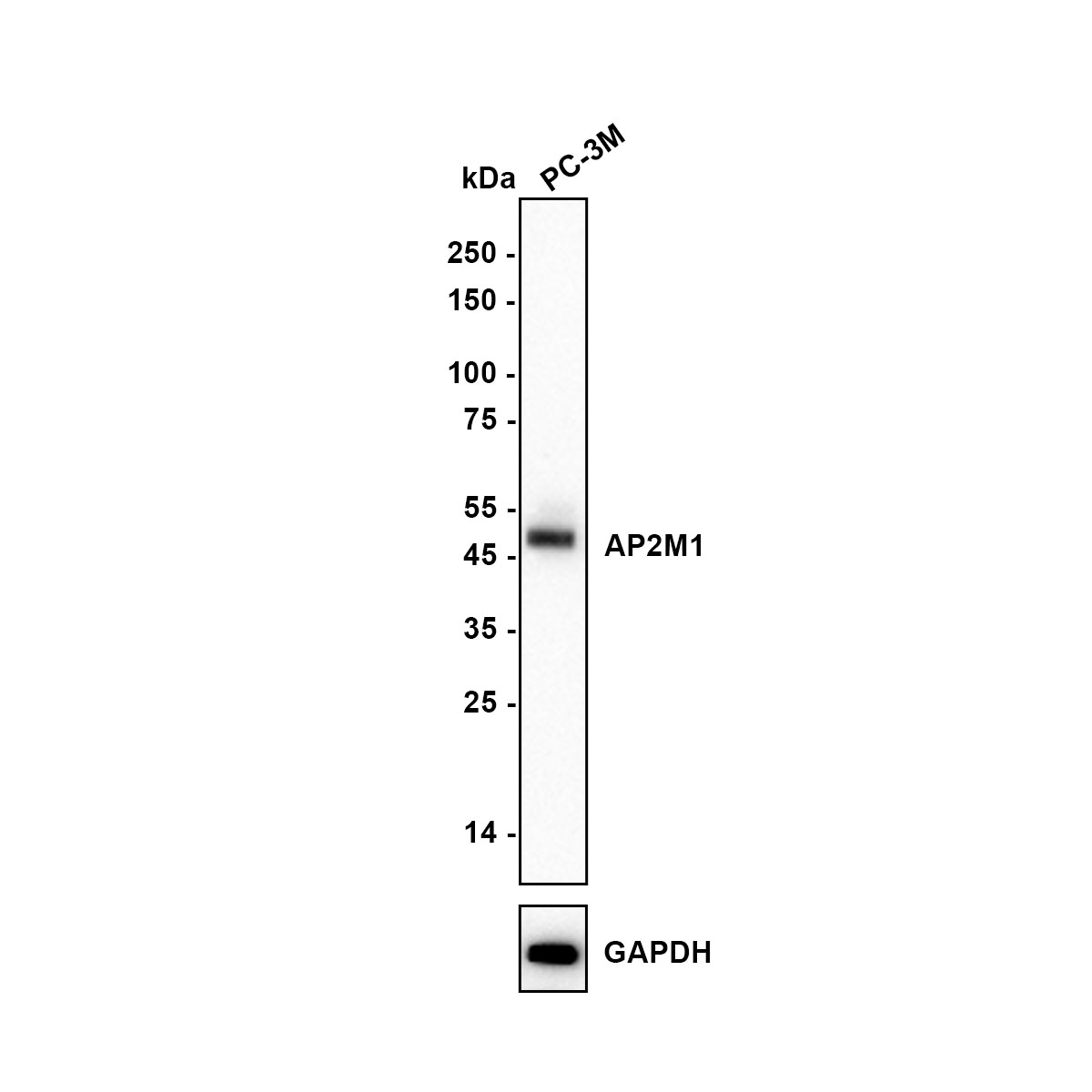
Western blot analysis of AP2M1 on PC-3M (Human prostate cancer cell) cell lysate with Rabbit anti-AP2M1 antibody at 1/5,000 dilution. Lysates/proteins at 10 µg/Lane. Exposure time: 20 seconds; Blocking: 5% NFDM/TBST, 1 hour at room temperature Primary antibody:1/5,000 in primary antibody dilution buffer, 2 hours at room temperature Secondary antibody: Goat anti-Rabbit IgG-HRP, 1/50,000 in 5% NFDM/TBST, 1 hour at room temperature.ICC/IF
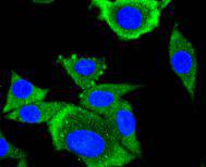
ICC staining AP2M1 in SH-SY-5Y cells (green). The nuclear counter stain is DAPI (blue). Cells were fixed in paraformaldehyde, permeabilised with 0.25% Triton X100/PBS.ICC/IF
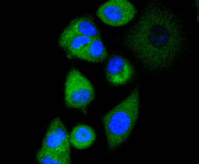
ICC staining AP2M1 in MCF-7 cells (green). The nuclear counter stain is DAPI (blue). Cells were fixed in paraformaldehyde, permeabilised with 0.25% Triton X100/PBS.IHC
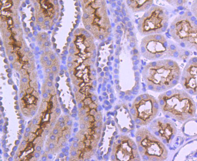
Immunohistochemical analysis of paraffin-embedded human kidney tissue using anti-AP2M1 antibody. Counter stained with hematoxylin.IHC
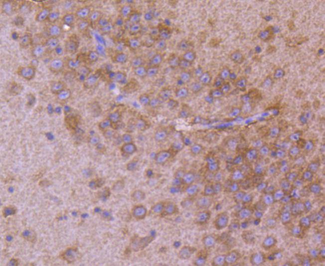
Immunohistochemical analysis of paraffin-embedded mouse brain tissue using anti-AP2M1 antibody. Counter stained with hematoxylin.FC
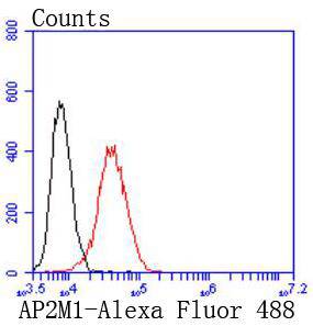
Flow cytometric analysis of PANC-1 cells with AP2M1 antibody at 1/50 dilution (red) compared with an unlabelled control (cells without incubation with primary antibody; black). Alexa Fluor 488-conjugated goat anti rabbit IgG was used as the secondary antibody| Product Name | AP2M1 Recombinant Rabbit Monoclonal Antibody |
|---|---|
| Antibody Type | Primary Antibodies |
| Product description | Adaptins are heterotetrameric subunits of adaptors, which are complexes involved in the formation of Clathrin-coated pits for vesicle-mediated endocytosis. Clathrin and its associated heterotetrameric protein complexes make up the main protein components of the coat surrounding the cytoplasmic face of coated vesicles. The Adaptin family, comprising a, b, b' and g classes, is also responsible for the transport of ligand-receptor complexes from plasma membranes and the trans-Golgi network to lysosomes. Two main types of adaptor proteins (APs), AP-1 and AP-2, are found in Clathrin-coated structures located at the Golgi complex and the plasma membrane of mammalian cells, respectively. Adaptor protein complex 2 (AP-2) is composed of two large Adaptins (a1A/AP2A1 and b1/AP2B1), a medium Adaptin (m2/AP-2m1) and a small Adaptin (s2 long/AP2S1). AP-2m1, a 435 amino acid protein, links Clathrin to receptors in coated vesicles. |
| Immunogen | Synthetic peptide within Human AP2M1 aa 351-400 / 435. |
| Clonality | Monoclonal |
|---|---|
| Isotype | IgG |
| Host Species | Recombinant rabbit |
| Tested Applications | FCICC/IFIHCWB |
| WB:1:500-1:5000 ICC/IF:1:50-1:200 IHC:1:50-1:200 FC:1:50-1:100 |
|
| Species Reactivity | HumanMouseRat |
| Concentration | 1mg/ml |
| Purification | Protein A |
| Alternative Names | Adapter-related protein complex 2 mu subunit antibody Adaptin mu 1 antibody Adaptin-mu2 antibody Adaptor protein complex AP 2 subunit mu antibody Adaptor protein complex AP-2 subunit mu antibody Adaptor related protein complex 2 mu 1 subunit antibody AP 2 mu 2 chain antibody AP-2 complex subunit mu antibody AP-2 mu chain antibody Ap2m1 antibody AP2M1_HUMAN antibody AP50 antibody CLAPM1 antibody Clathrin adaptor complex AP2 mu subunit antibody Clathrin assembly protein complex 2 medium chain antibody Clathrin associated/assembly/adaptor protein medium 1 antibody Clathrin coat adaptor protein AP50 antibody Clathrin coat assembly protein AP50 antibody Clathrin coat associated protein AP50 antibody Clathrin coat-associated protein AP50 antibody HA2 50 kDa subunit antibody mu2 antibody Plasma membrane adaptor AP-2 50 kDa protein antibody |
|---|---|
| Molecular Weight(MW) | 50 kDa |
| Cellular Localization | Cell membrane, Membrane. |
WB
Western blot analysis of AP2M1 on PC-3M (Human prostate cancer cell) cell lysate with Rabbit anti-AP2M1 antibody at 1/5,000 dilution. Lysates/proteins at 10 µg/Lane. Exposure time: 20 seconds; Blocking: 5% NFDM/TBST, 1 hour at room temperature Primary antibody:1/5,000 in primary antibody dilution buffer, 2 hours at room temperature Secondary antibody: Goat anti-Rabbit IgG-HRP, 1/50,000 in 5% NFDM/TBST, 1 hour at room temperature.
ICC/IF
ICC staining AP2M1 in SH-SY-5Y cells (green). The nuclear counter stain is DAPI (blue). Cells were fixed in paraformaldehyde, permeabilised with 0.25% Triton X100/PBS.
ICC/IF
ICC staining AP2M1 in MCF-7 cells (green). The nuclear counter stain is DAPI (blue). Cells were fixed in paraformaldehyde, permeabilised with 0.25% Triton X100/PBS.
IHC
Immunohistochemical analysis of paraffin-embedded human kidney tissue using anti-AP2M1 antibody. Counter stained with hematoxylin.
IHC
Immunohistochemical analysis of paraffin-embedded mouse brain tissue using anti-AP2M1 antibody. Counter stained with hematoxylin.
FC
Flow cytometric analysis of PANC-1 cells with AP2M1 antibody at 1/50 dilution (red) compared with an unlabelled control (cells without incubation with primary antibody; black). Alexa Fluor 488-conjugated goat anti rabbit IgG was used as the secondary antibody| Application Notes | WB:1:500-1:5000 ICC/IF:1:50-1:200 IHC:1:50-1:200 FC:1:50-1:100 |
|---|
| Form | Liquid |
|---|---|
| Storage Instructions | Store at +4℃ after thawing. Aliquot store at -20℃ or -80℃. Avoid repeated freeze / thaw cycles. |
| Storage Buffer | 1*TBS (pH7.4), 1%BSA, 40%Glycerol. Preservative: 0.05% Sodium Azide. |


