Application
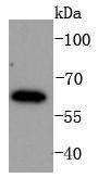
Fig1: Western blot analysis of IRF6 on K562 cells lysates using anti-IRF6 antibody at 1/1,000 dilution.Application
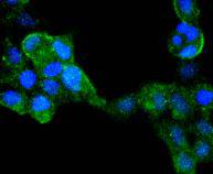
Fig2: ICC staining IRF6 in 293 cells (green). The nuclear counter stain is DAPI (blue). Cells were fixed in paraformaldehyde, permeabilised with 0.25% Triton X100/PBS.Application
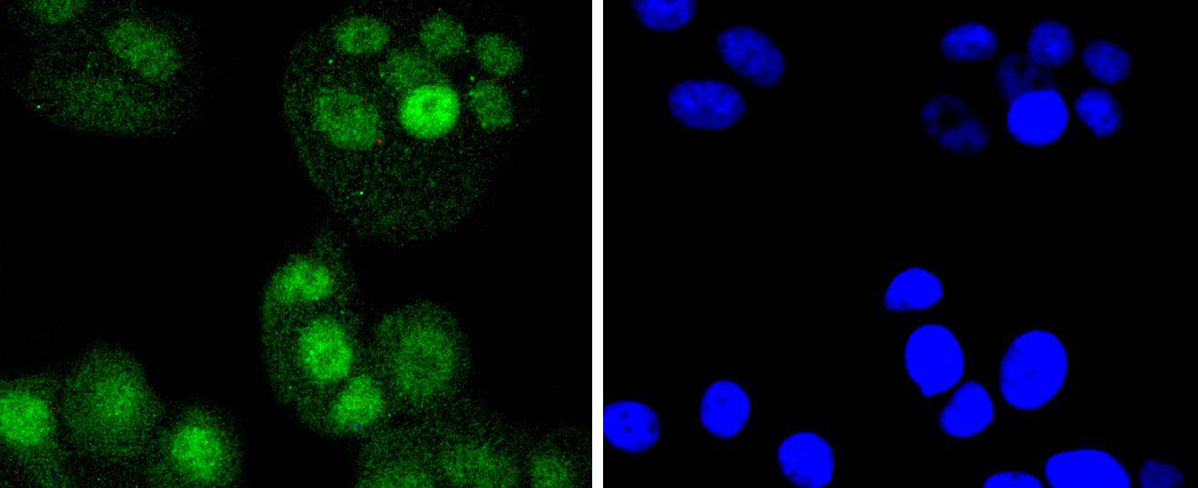
Fig3: ICC staining IRF6 in MCF-7 cells (green). The nuclear counter stain is DAPI (blue). Cells were fixed in paraformaldehyde, permeabilised with 0.25% Triton X100/PBS.Application
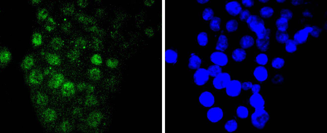
Fig4: ICC staining IRF6 in SW480 cells (green). The nuclear counter stain is DAPI (blue). Cells were fixed in paraformaldehyde, permeabilised with 0.25% Triton X100/PBS.Application
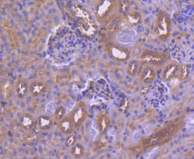
Fig5: Immunohistochemical analysis of paraffin-embedded mouse kidney tissue using anti-IRF6 antibody. Counter stained with hematoxylin.Application
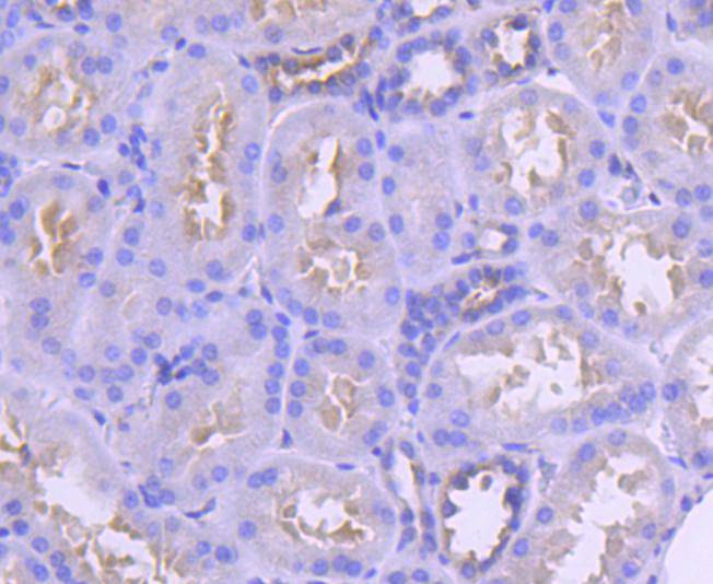
Fig6: Immunohistochemical analysis of paraffin-embedded human kidney tissue using anti-IRF6 antibody. Counter stained with hematoxylin.| Product Name | IRF6 [SD202-6] |
|---|---|
| Antibody Type | Primary Antibodies |
| Antigen Alias | Interferon regulatory factor 6 antibody IRF 6 antibody IRF-6 antibody IRF6 antibody IRF6_HUMAN antibody LPS antibody OFC 6 antibody OFC6 antibody OTTHUMP00000034677 antibody OTTHUMP00000034678 antibody PIT antibody PPS antibody PPS1 antibody VWS antibody VWS1 antibody |
| Product description | Interferon regulatory factor-1 (IRF-1) and IRF-2 have been identified as novel DNA-binding factors that function as regulators of both type I interferon (interferon-α and β) and interferon-inducible genes. The two factors are structurally related, particularly in their N-terminal regions, which confer DNA binding specificity. In addition, both bind to the same sequence within the promoters of interferon-α and interferon-β genes. IRF-1 functions as an activator of interferon transcription, while IRF-2 binds to the same cis elements and represses IRF-1 action. IRF-1 and IRF-2 have been reported to act in a mutually antagonistic manner in regulating cell growth; overexpression of the repressor IRF-2 leads to cell transformation while concomitant overexpression of IRF-1 causes reversion. IRF-1 and IRF-2 are members of a larger family of DNA binding proteins that includes IRF-3, IRF-4, IRF-5, IRF-6, IRF-7, ISGF-3γ p48 and IFN consensus sequence-binding protein (ICSBP). |
| Immunogen | recombinant protein |
| Clonality | Monoclonal |
|---|---|
| Isotype | IgG |
| Host Species | Recombinant rabbit |
| Tested Applications | WBICC/IFIHC |
| WB:1:1,000-1:2,000 ICC:1:50-1:200 IHC:1:50-1:200 |
|
| Species Reactivity | HumanMouseRat |
| Concentration | 1mg/ml |
| Alternative Names | Interferon regulatory factor 6 antibody IRF 6 antibody IRF-6 antibody IRF6 antibody IRF6_HUMAN antibody LPS antibody OFC 6 antibody OFC6 antibody OTTHUMP00000034677 antibody OTTHUMP00000034678 antibody PIT antibody PPS antibody PPS1 antibody VWS antibody VWS1 antibody |
|---|---|
| Molecular Weight(MW) | 60 kDa |
| Cellular Localization | Nucleus, Cytoplasm. |
Application
Fig1: Western blot analysis of IRF6 on K562 cells lysates using anti-IRF6 antibody at 1/1,000 dilution.
Application
Fig2: ICC staining IRF6 in 293 cells (green). The nuclear counter stain is DAPI (blue). Cells were fixed in paraformaldehyde, permeabilised with 0.25% Triton X100/PBS.
Application
Fig3: ICC staining IRF6 in MCF-7 cells (green). The nuclear counter stain is DAPI (blue). Cells were fixed in paraformaldehyde, permeabilised with 0.25% Triton X100/PBS.
Application
Fig4: ICC staining IRF6 in SW480 cells (green). The nuclear counter stain is DAPI (blue). Cells were fixed in paraformaldehyde, permeabilised with 0.25% Triton X100/PBS.
Application
Fig5: Immunohistochemical analysis of paraffin-embedded mouse kidney tissue using anti-IRF6 antibody. Counter stained with hematoxylin.
Application
Fig6: Immunohistochemical analysis of paraffin-embedded human kidney tissue using anti-IRF6 antibody. Counter stained with hematoxylin.| Positive Control | K562, MCF-7, 293, SW480, human kidney tissue, mouse kidney tissue. |
|---|---|
| Application Notes | WB:1:1,000-1:2,000 ICC:1:50-1:200 IHC:1:50-1:200 |
| Form | Liquid |
|---|---|
| Storage Instructions | Store at +4℃ after thawing. Aliquot store at -20℃ or -80℃. Avoid repeated freeze / thaw cycles. |
| Storage Buffer | 1*TBS (pH7.4), 1%BSA, 40%Glycerol. Preservative: 0.05% Sodium Azide. |


