Application
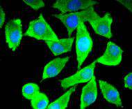
Fig1: ICC staining Tau in SH-SY-5Y cells (green). The nuclear counter stain is DAPI (blue). Cells were fixed in paraformaldehyde, permeabilised with 0.25% Triton X100/PBS.Application
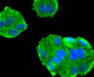
Fig2: ICC staining Tau in Hela cells (green). The nuclear counter stain is DAPI (blue). Cells were fixed in paraformaldehyde, permeabilised with 0.25% Triton X100/PBS.Application
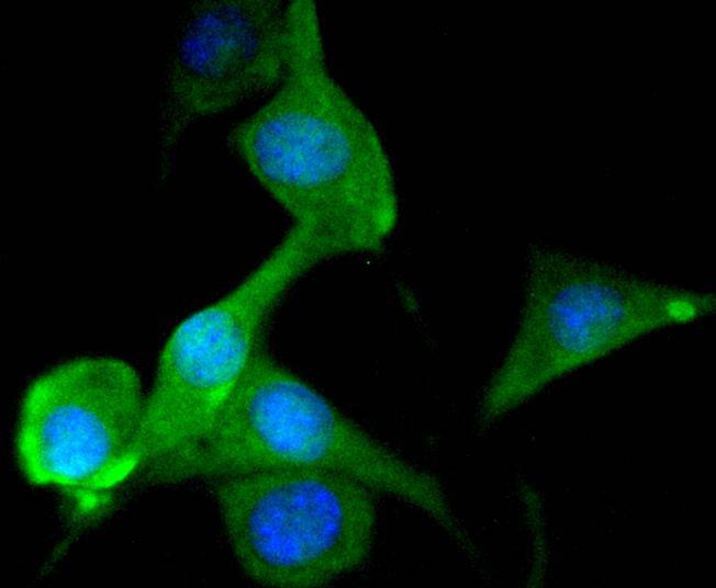
Fig3: ICC staining Tau in SHG-44 cells (green). The nuclear counter stain is DAPI (blue). Cells were fixed in paraformaldehyde, permeabilised with 0.25% Triton X100/PBS.Application
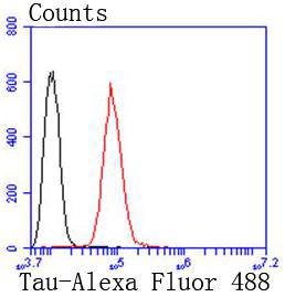
Fig4: Flow cytometric analysis of SH-SY-5Y cells with Tau antibody at 1/50 dilution (red) compared with an unlabelled control (cells without incubation with primary antibody; black). Alexa Fluor 488-conjugated goat anti rabbit IgG was used as the secondary antibody| Product Name | Tau [SD205-09] |
|---|---|
| Antibody Type | Primary Antibodies |
| Antigen Alias | AI413597 antibody AW045860 antibody DDPAC antibody FLJ31424 antibody FTDP 17 antibody G protein beta1/gamma2 subunit interacting factor 1 antibody MAPT antibody MAPTL antibody MGC134287 antibody MGC138549 antibody MGC156663 antibody Microtubule associated protein tau antibody Microtubule associated protein tau isoform 4 antibody Microtubule-associated protein tau antibody MSTD antibody Mtapt antibody MTBT1 antibody MTBT2 antibody Neurofibrillary tangle protein antibody Paired helical filament tau antibody Paired helical filament-tau antibody PHF tau antibody PHF-tau antibody PPND antibody PPP1R103 antibody Protein phosphatase 1, regulatory subunit 103 antibody pTau antibody RNPTAU antibody TAU antibody TAU_HUMAN antibody Tauopathy and respiratory failure, included antibody |
| Product description | Tau, also known as MAPT (microtubule-associated protein tau), MAPTL, MTBT1 or TAU, is a 758 amino acid protein that localizes to the cytoplasm, as well as to the cytoskeleton and the cell membrane, and contains four Tau/MAP repeats. Expressed in neuronal tissue and existing as multiple alternatively spliced isoforms, Tau functions to promote microtubule assembly and stability and is thought to be involved in the maintenance of neuronal polarity. Tau may also link microtubules with neural plasma membrane components and, addition to its role in microtubule stability, is also necessary for cytoskeletal plasticity. Tau is highly subject to a variety of post-translational modifications, including phosphorylation on serine and threonine residues, polyubiquitination (and subsequent proteasomal degradation) and glycation of specific Tau isoforms. Defects in the gene encoding Tau are associated with Alzheimers disease, pallido-ponto-nigral degeneration (PPND), corticobasal degeneration (CBD) and progressive supranuclear palsy (PSP). |
| Immunogen | recombinant protein |
| Clonality | Monoclonal |
|---|---|
| Isotype | IgG |
| Host Species | Recombinant rabbit |
| Tested Applications | WBICC/IFFC |
| WB:1:1,000 ICC:1:100-1:500 FC:1:50-1:100 |
|
| Species Reactivity | HumanMouseRat |
| Concentration | 1mg/ml |
| Alternative Names | AI413597 antibody
AW045860 antibody
DDPAC antibody
FLJ31424 antibody
FTDP 17 antibody
G protein beta1/gamma2 subunit interacting factor 1 antibody
MAPT antibody
MAPTL antibody
MGC134287 antibody
MGC138549 antibody
MGC156663 antibody
Microtubule associated protein tau antibody
Microtubule associated protein tau isoform 4 antibody
Microtubule-associated protein tau antibody
MSTD antibody
Mtapt antibody
MTBT1 antibody
MTBT2 antibody
Neurofibrillary tangle protein antibody
Paired helical filament tau antibody
Paired helical filament-tau antibody
PHF tau antibody
PHF-tau antibody
PPND antibody
PPP1R103 antibody
Protein phosphatase 1 regulatory subunit 103 antibody pTau antibody RNPTAU antibody TAU antibody TAU_HUMAN antibody Tauopathy and respiratory failure included antibody |
|---|---|
| Molecular Weight(MW) | 79 kDa |
| Cellular Localization | Cytoplasm, Cell membrane, Cell projection. |
Application
Fig1: ICC staining Tau in SH-SY-5Y cells (green). The nuclear counter stain is DAPI (blue). Cells were fixed in paraformaldehyde, permeabilised with 0.25% Triton X100/PBS.
Application
Fig2: ICC staining Tau in Hela cells (green). The nuclear counter stain is DAPI (blue). Cells were fixed in paraformaldehyde, permeabilised with 0.25% Triton X100/PBS.
Application
Fig3: ICC staining Tau in SHG-44 cells (green). The nuclear counter stain is DAPI (blue). Cells were fixed in paraformaldehyde, permeabilised with 0.25% Triton X100/PBS.
Application
Fig4: Flow cytometric analysis of SH-SY-5Y cells with Tau antibody at 1/50 dilution (red) compared with an unlabelled control (cells without incubation with primary antibody; black). Alexa Fluor 488-conjugated goat anti rabbit IgG was used as the secondary antibody| Positive Control | SH-SY-5Y, Hela, SHG-44. |
|---|---|
| Application Notes | WB:1:1,000 ICC:1:100-1:500 FC:1:50-1:100 |
| Form | Liquid |
|---|---|
| Storage Instructions | Store at +4℃ after thawing. Aliquot store at -20℃ or -80℃. Avoid repeated freeze / thaw cycles. |
| Storage Buffer | 1*TBS (pH7.4), 1%BSA, 40%Glycerol. Preservative: 0.05% Sodium Azide. |


