Application
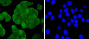
Fig1: ICC staining IRS1 in SW480 cells (green). The nuclear counter stain is DAPI (blue). Cells were fixed in paraformaldehyde, permeabilised with 0.25% Triton X100/PBS.Application
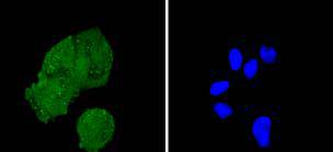
Fig2: ICC staining IRS1 in Hela cells (green). The nuclear counter stain is DAPI (blue). Cells were fixed in paraformaldehyde, permeabilised with 0.25% Triton X100/PBS.Application
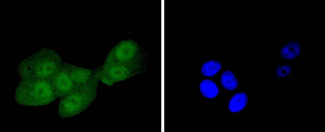
Fig3: ICC staining IRS1 in MCF-7 cells (green). The nuclear counter stain is DAPI (blue). Cells were fixed in paraformaldehyde, permeabilised with 0.25% Triton X100/PBS.Application
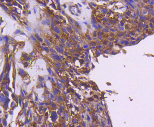
Fig4: Immunohistochemical analysis of paraffin-embedded human breast carcinoma tissue using anti-IRS1 antibody. Counter stained with hematoxylin.Application
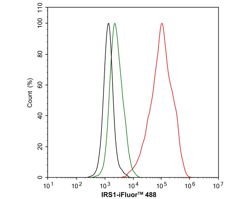
Fig5: Flow cytometric analysis of HeLa cells labeling IRS1. Cells were fixed and permeabilized. Then stained with the primary antibody (1μg/mL) (red) compared with Rabbit IgG Isotype Control (green). After incubation of the primary antibody at +4℃ for an hour, the cells were stained with a iFluor™ 488 conjugate-Goat anti-Rabbit IgG Secondary antibody at 1/1,000 dilution for 30 minutes at +4℃. Unlabelled sample was used as a control (cells without incubation with primary antibody; black).| Product Name | IRS1 Recombinant Rabbit Monoclonal Antibody |
|---|---|
| Antibody Type | Primary Antibodies |
| Product description | The insulin receptor substrate-1 (IRS-1), a protein major substrate of the insulin receptor, is phosphorylated in response to stimulation of cells by insulin, insulin-like growth factor 1 (IGF-1) and interleukin 4 (IL-4). IRS-1 is phosphorylated on serine, threonine and tyrosine residues in a variety of tissues. An insulin-sensitive serine/threonine kinase casein kinase II mediates a portion of the insulin-stimulated serine/threonine phosphorylation of overexpressed IRS-1 in vivo. Thr 502 is identified as the major casein kinase II-catalyzed phosphorylation site in rat IRS-1, and Ser 99 is an additional phosphorylation site catalyzed by casein kinase II. Thus, casein kinase II-catalyzed phosphorylation of IRS-1 may be a component of the intracellular insulin signaling cascade. IRS-1 contains three putative binding sites for 14-3-3 (Ser 270, Ser 374 and Ser 641) and the motif around Ser 270 is located in the phosphotyrosine binding domain of IRS-1, which is responsible for the interaction with the insulin receptor. The association of 14-3-3 with IRS-1 increases significantly upon treatment with okadaic acid, a potent serine/ threonine phosphatase inhibitor. Therefore, the association of 14-3-3 protein may play a role in the regulation of insulin sensitivity by interrupting the association between the insulin receptor and IRS-1. |
| Immunogen | Synthetic peptide within human IRS1 aa 1160-1242. |
| Clonality | monoclonal |
|---|---|
| Isotype | IgG |
| Host Species | Recombinant rabbit |
| Tested Applications | FCICC/IFIHCWB |
| WB:1:1,000-1:2,000 ICC:1:50-1:200 IHC:1:50-1:200 FC:1:1000 |
|
| Species Reactivity | Human |
| Concentration | 1mg/ml |
| Purification | Protein A |
| Alternative Names | HIRS 1 antibody HIRS1 antibody Insulin receptor substrate 1 antibody IRS 1 antibody IRS-1 antibody IRS1 antibody IRS1_HUMAN antibody OTTHUMP00000164234 antibody |
|---|---|
| Molecular Weight(MW) | 170 kDa |
| Cellular Localization | Cytoplasm,Nucleus. |
| SwissProt ID | P35568 |
|---|
Application
Fig1: ICC staining IRS1 in SW480 cells (green). The nuclear counter stain is DAPI (blue). Cells were fixed in paraformaldehyde, permeabilised with 0.25% Triton X100/PBS.
Application
Fig2: ICC staining IRS1 in Hela cells (green). The nuclear counter stain is DAPI (blue). Cells were fixed in paraformaldehyde, permeabilised with 0.25% Triton X100/PBS.
Application
Fig3: ICC staining IRS1 in MCF-7 cells (green). The nuclear counter stain is DAPI (blue). Cells were fixed in paraformaldehyde, permeabilised with 0.25% Triton X100/PBS.
Application
Fig4: Immunohistochemical analysis of paraffin-embedded human breast carcinoma tissue using anti-IRS1 antibody. Counter stained with hematoxylin.
Application
Fig5: Flow cytometric analysis of HeLa cells labeling IRS1. Cells were fixed and permeabilized. Then stained with the primary antibody (1μg/mL) (red) compared with Rabbit IgG Isotype Control (green). After incubation of the primary antibody at +4℃ for an hour, the cells were stained with a iFluor™ 488 conjugate-Goat anti-Rabbit IgG Secondary antibody at 1/1,000 dilution for 30 minutes at +4℃. Unlabelled sample was used as a control (cells without incubation with primary antibody; black).| Positive Control | SW480, MCF-7, Hela, human breast carcinoma tissue. |
|---|---|
| Application Notes | WB:1:1,000-1:2,000 ICC:1:50-1:200 IHC:1:50-1:200 FC:1:1000 |
| Form | Liquid |
|---|---|
| Storage Instructions | Store at +4℃ after thawing. Aliquot store at -20℃ or -80℃. Avoid repeated freeze / thaw cycles. |
| Storage Buffer | 1*TBS (pH7.4), 1%BSA, 40%Glycerol. Preservative: 0.05% Sodium Azide. |


