WB
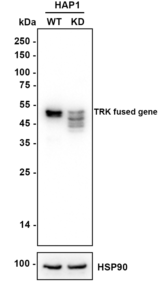
Western blot analysis of TRK fused gene on different lysates with Rabbit anti-TRK fused gene antibody at 1/1,000 dilution. Lane 1: HAP1-parental cell lysate, Lane 2: HAP1-TRK fused gene KD cell lysate, Lysates/proteins at 10 µg/Lane. Exposure time: 6 seconds; 4-20% SDS-PAGE gel. Proteins were transferred to a PVDF membrane and blocked with 5% NFDM/TBST for 1 hour at room temperature. The primary antibody at 1/1,000 dilution was used in primary antibody dilution at 4℃ overnight. Goat Anti-Rabbit IgG - HRP Secondary Antibody at 1/50,000 dilution was used for 1 hour at room temperature.ICC/IF
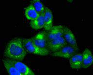
ICC staining TRK fused gene in Hela cells (green). The nuclear counter stain is DAPI (blue). Cells were fixed in paraformaldehyde, permeabilised with 0.25% Triton X100/PBS.ICC/IF
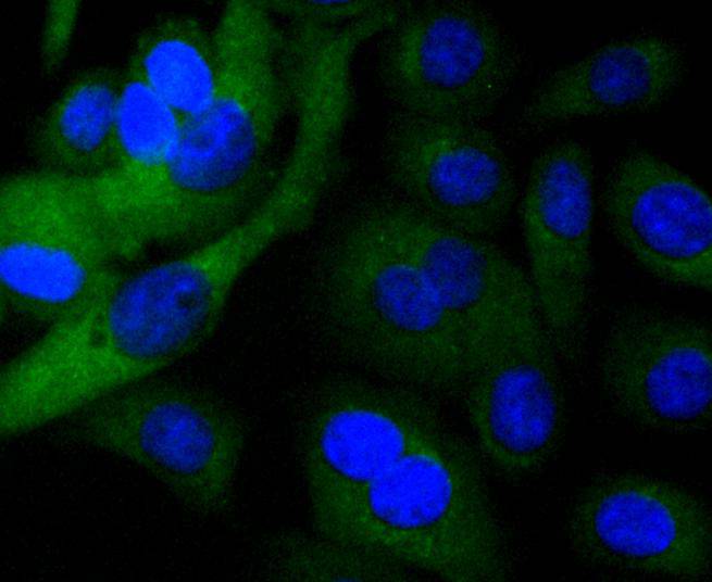
ICC staining TRK fused gene in SKOV-3 cells (green). The nuclear counter stain is DAPI (blue). Cells were fixed in paraformaldehyde, permeabilised with 0.25% Triton X100/PBS.ICC/IF
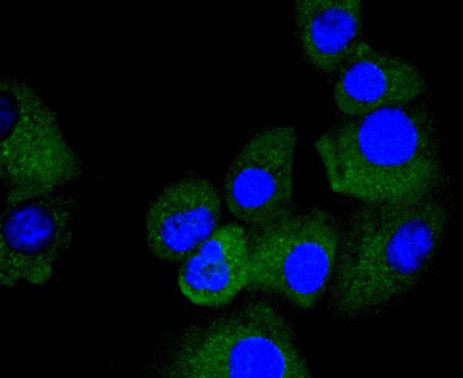
ICC staining TRK fused gene in A549 cells (green). The nuclear counter stain is DAPI (blue). Cells were fixed in paraformaldehyde, permeabilised with 0.25% Triton X100/PBS.IHC
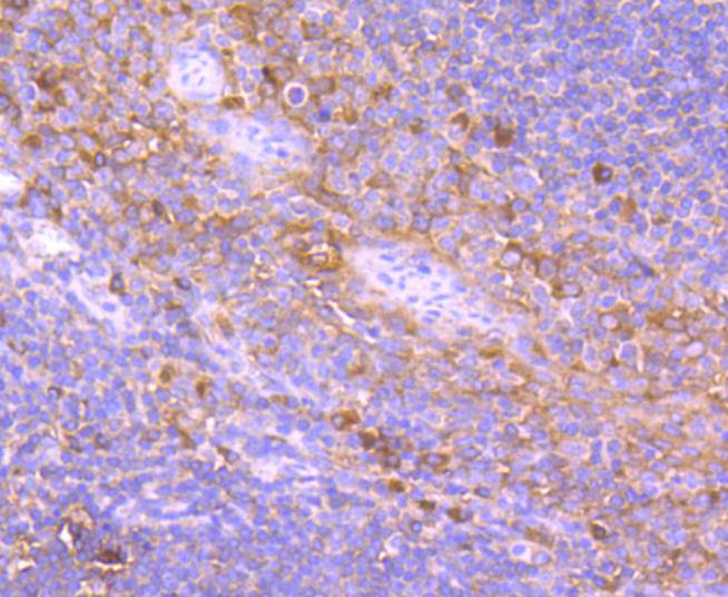
Immunohistochemical analysis of paraffin-embedded human tonsil tissue using anti-TRK fused gene antibody. Counter stained with hematoxylin.IHC
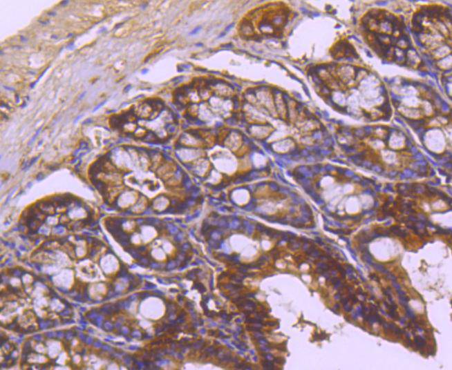
Immunohistochemical analysis of paraffin-embedded mouse colon tissue using anti-TRK fused gene antibody. Counter stained with hematoxylin.IHC
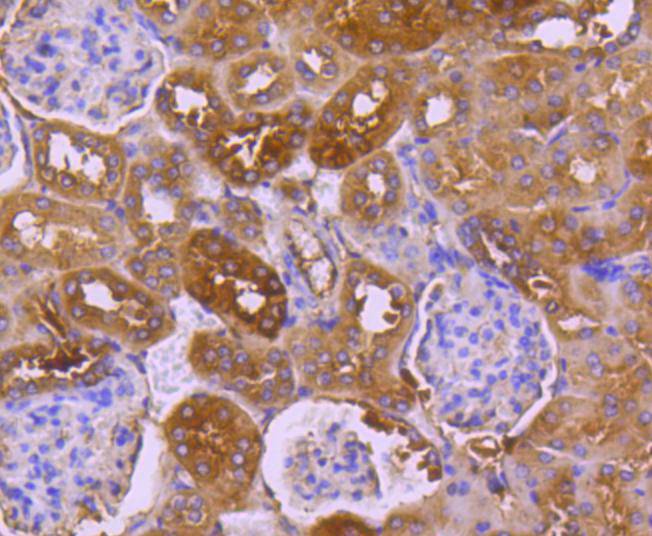
Immunohistochemical analysis of paraffin-embedded mouse kidney tissue using anti-TRK fused gene antibody. Counter stained with hematoxylin.IHC
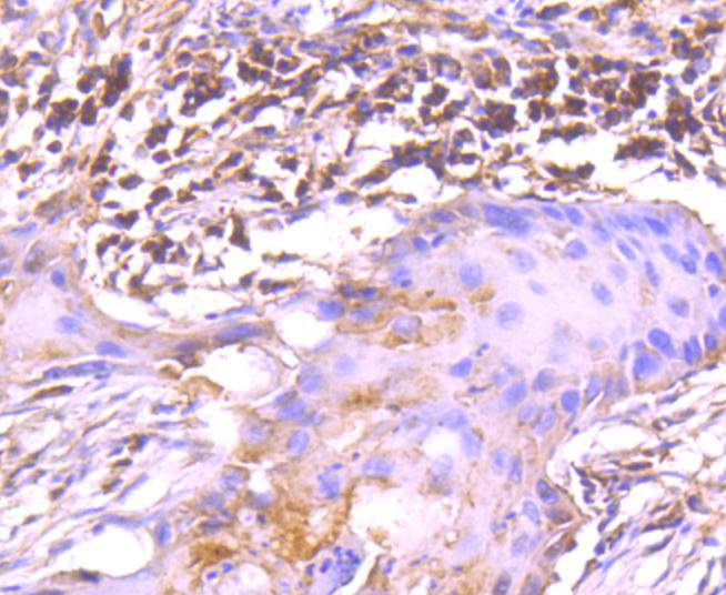
Immunohistochemical analysis of paraffin-embedded human lung cancer tissue using anti-TRK fused gene antibody. Counter stained with hematoxylin.IHC
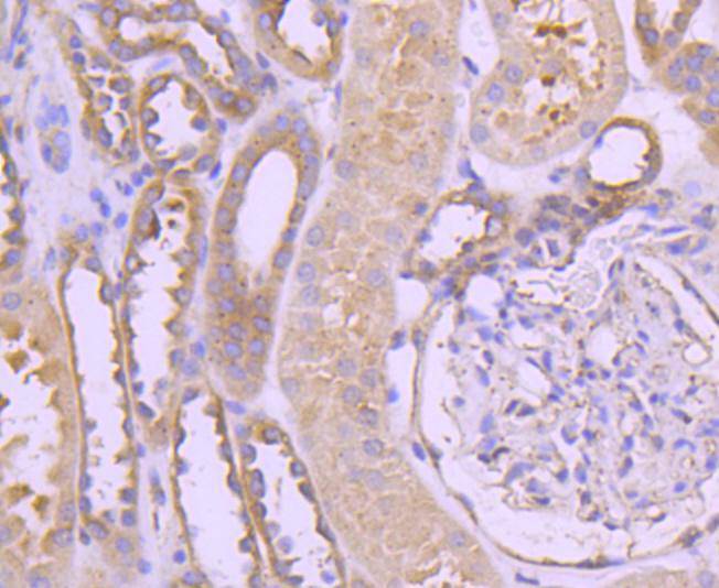
Immunohistochemical analysis of paraffin-embedded human kidney tissue using anti-TRK fused gene antibody. Counter stained with hematoxylin.| Product Name | TRK fused gene Recombinant Rabbit Monoclonal Antibody |
|---|---|
| Antibody Type | Primary Antibodies |
| Product description | Oncogenic rearrangements of the NTRK1 gene, which encodes the Trk A protein, are frequently detected in thyroid carcinomas. Such rearrangements fuse the NTRK1 tyrosine kinase domain to 5'-end sequences of different genes. TRK-T3 contains 1,412 nucleotides of NTRK1 preceded by 598 nucleotides belonging to TFG (TRK-fused gene), a ubiquitously expressed gene located on chromosome 3. The TRK-T3 protein within the TFG region contains a coiled-coil motif that gives the oncoprotein the capability to form complexes. The cytoplasmic TRK-T3 protein binds to and phosphorylates the Shc and SNT1/FRS2 adaptor proteins, both of which are involved in coupling the receptor tyrosine kinase to the mitogen-activated protein kinase pathway by recruiting Grb2/SOS. SHP-1 also interacts with and down-regulates TRK-T3. |
| Immunogen | Synthetic peptide within Human TRK aa 351-400 / 400. |
| Clonality | Monoclonal |
|---|---|
| Isotype | IgG |
| Host Species | Recombinant rabbit |
| Tested Applications | ICC/IFIHCWB |
| WB:1:500-1:2000 IHC:1:50-1:200 ICC/IF:1:50-1:200 |
|
| Species Reactivity | HumanMouseRat |
| Concentration | 1mg/ml |
| Purification | Protein A |
| Alternative Names | FLJ36137 antibody HMSNP antibody OTTHUMP00000214045 antibody OTTHUMP00000214046 antibody OTTHUMP00000214047 antibody OTTHUMP00000214048 antibody Protein TFG antibody SPG57 antibody TF6 antibody TFG antibody TFG_HUMAN antibody TRK fused antibody TRK fused gene antibody TRK fused gene protein antibody TRK-fused gene protein antibody TRKT3 antibody TRKT3 oncogene antibody |
|---|---|
| Molecular Weight(MW) | 43 kDa(Observed band size: 50 kDa) |
| Cellular Localization | Cytoplasm. |
WB
Western blot analysis of TRK fused gene on different lysates with Rabbit anti-TRK fused gene antibody at 1/1,000 dilution. Lane 1: HAP1-parental cell lysate, Lane 2: HAP1-TRK fused gene KD cell lysate, Lysates/proteins at 10 µg/Lane. Exposure time: 6 seconds; 4-20% SDS-PAGE gel. Proteins were transferred to a PVDF membrane and blocked with 5% NFDM/TBST for 1 hour at room temperature. The primary antibody at 1/1,000 dilution was used in primary antibody dilution at 4℃ overnight. Goat Anti-Rabbit IgG - HRP Secondary Antibody at 1/50,000 dilution was used for 1 hour at room temperature.
ICC/IF
ICC staining TRK fused gene in Hela cells (green). The nuclear counter stain is DAPI (blue). Cells were fixed in paraformaldehyde, permeabilised with 0.25% Triton X100/PBS.
ICC/IF
ICC staining TRK fused gene in SKOV-3 cells (green). The nuclear counter stain is DAPI (blue). Cells were fixed in paraformaldehyde, permeabilised with 0.25% Triton X100/PBS.
ICC/IF
ICC staining TRK fused gene in A549 cells (green). The nuclear counter stain is DAPI (blue). Cells were fixed in paraformaldehyde, permeabilised with 0.25% Triton X100/PBS.
IHC
Immunohistochemical analysis of paraffin-embedded human tonsil tissue using anti-TRK fused gene antibody. Counter stained with hematoxylin.
IHC
Immunohistochemical analysis of paraffin-embedded mouse colon tissue using anti-TRK fused gene antibody. Counter stained with hematoxylin.
IHC
Immunohistochemical analysis of paraffin-embedded mouse kidney tissue using anti-TRK fused gene antibody. Counter stained with hematoxylin.
IHC
Immunohistochemical analysis of paraffin-embedded human lung cancer tissue using anti-TRK fused gene antibody. Counter stained with hematoxylin.
IHC
Immunohistochemical analysis of paraffin-embedded human kidney tissue using anti-TRK fused gene antibody. Counter stained with hematoxylin.| Application Notes | WB:1:500-1:2000 IHC:1:50-1:200 ICC/IF:1:50-1:200 |
|---|
| Form | Liquid |
|---|---|
| Storage Instructions | Store at +4℃ after thawing. Aliquot store at -20℃ or -80℃. Avoid repeated freeze / thaw cycles. |
| Storage Buffer | 1*TBS (pH7.4), 1%BSA, 40%Glycerol. Preservative: 0.05% Sodium Azide. |


