Application
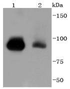
Fig1: Western blot analysis of STAT5b on different lysates using anti-STAT5b antibody at 1/1,000 dilution. Positive control: Lane 1: K562 Lane 2: HelaApplication
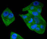
Fig2: ICC staining STAT5b in Hela cells (green). The nuclear counter stain is DAPI (blue). Cells were fixed in paraformaldehyde, permeabilised with 0.25% Triton X100/PBS.Application
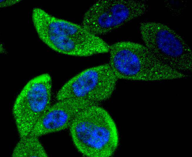
Fig3: ICC staining STAT5b in PC-3M cells (green). The nuclear counter stain is DAPI (blue). Cells were fixed in paraformaldehyde, permeabilised with 0.25% Triton X100/PBS.Application
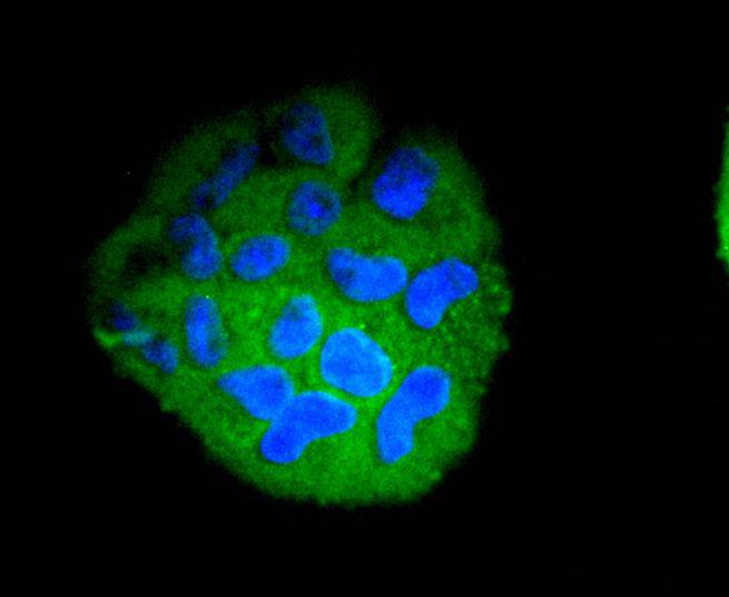
Fig4: ICC staining STAT5b in RH-35 cells (green). The nuclear counter stain is DAPI (blue). Cells were fixed in paraformaldehyde, permeabilised with 0.25% Triton X100/PBS.Application
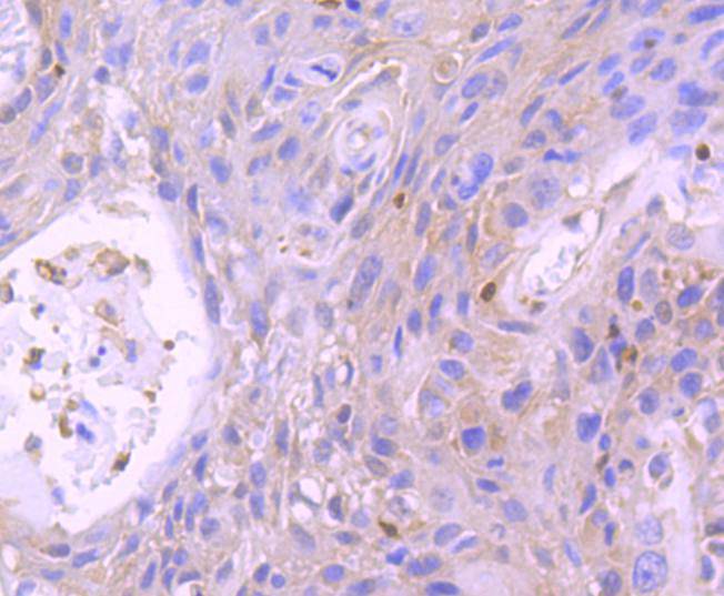
Fig5: Immunohistochemical analysis of paraffin-embedded human breast carcinoma tissue using anti-STAT5b antibody. Counter stained with hematoxylin.Application
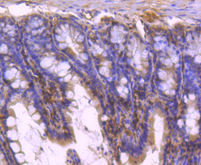
Fig6: Immunohistochemical analysis of paraffin-embedded mouse colon tissue using anti-STAT5b antibody. Counter stained with hematoxylin.Application
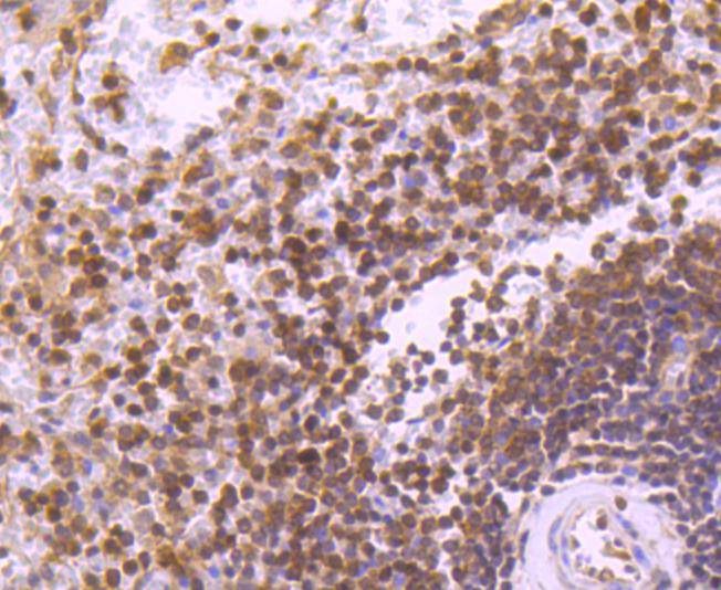
Fig7: Immunohistochemical analysis of paraffin-embedded human spleen tissue using anti-STAT5b antibody. Counter stained with hematoxylin.Application
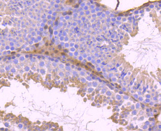
Fig8: Immunohistochemical analysis of paraffin-embedded mouse testis tissue using anti-STAT5b antibody. Counter stained with hematoxylin.| Product Name | STAT5b [SD08-08] |
|---|---|
| Antibody Type | Primary Antibodies |
| Antigen Alias | Signal transducer and activator of transcription 5B antibody STA5B_HUMAN antibody STAT5 antibody Stat5b antibody Transcription factor STAT5B antibody |
| Product description | Signal transducer and activator of transcription 5A (Stat5a) and Stat5b, which share 96% homology, undergo receptor tyrosine kinase or G protein-coupled receptor-dependent phosphorylation in response to cytokines or growth factors, and then form homo- or heterodimers that translocate to the nucleus, where they initiate transcription. Activation of Stat5a via IL-2, IL-3, IL-7/ GM-CSF, erythropoietin, thrombopoietin and growth hormones influences proliferation, differentiation and apoptosis in lymphohematopoietic cells. Phos-phorylation of Stat5a at Ser127/Ser128 and Ser779 are contigent on ErbB-4-mediated activation of Stat5a. Activation of Stat5b via IL-2, IL-4, CSF-1 and growth hormones influences TCR signaling, apoptosis, adult mammary gland development and sexual dimorphism of liver gene expression. Stat5b is the major liver-expressed Stat5 form that has been shown to fuse with the retin-oic acid receptor a gene in acute promyelocytic leukemias (APLL). Stat5a/b null mice have severely impaired lymphoid development and differentiation. |
| Immunogen | recombinant protein |
| Clonality | Monoclonal |
|---|---|
| Isotype | IgG |
| Host Species | Recombinant rabbit |
| Tested Applications | WBICC/IFIHCIP |
| WB:1:1,000-1:2,000 ICC:1:100-1:500 IHC:1:50-1:200 |
|
| Species Reactivity | HumanMouseRat |
| Concentration | 1mg/ml |
| Alternative Names | Signal transducer and activator of transcription 5B antibody STA5B_HUMAN antibody STAT5 antibody Stat5b antibody Transcription factor STAT5B antibody |
|---|---|
| Molecular Weight(MW) | 90 kDa |
| Cellular Localization | Cytoplasm, Nucleus. |
Application
Fig1: Western blot analysis of STAT5b on different lysates using anti-STAT5b antibody at 1/1,000 dilution. Positive control: Lane 1: K562 Lane 2: Hela
Application
Fig2: ICC staining STAT5b in Hela cells (green). The nuclear counter stain is DAPI (blue). Cells were fixed in paraformaldehyde, permeabilised with 0.25% Triton X100/PBS.
Application
Fig3: ICC staining STAT5b in PC-3M cells (green). The nuclear counter stain is DAPI (blue). Cells were fixed in paraformaldehyde, permeabilised with 0.25% Triton X100/PBS.
Application
Fig4: ICC staining STAT5b in RH-35 cells (green). The nuclear counter stain is DAPI (blue). Cells were fixed in paraformaldehyde, permeabilised with 0.25% Triton X100/PBS.
Application
Fig5: Immunohistochemical analysis of paraffin-embedded human breast carcinoma tissue using anti-STAT5b antibody. Counter stained with hematoxylin.
Application
Fig6: Immunohistochemical analysis of paraffin-embedded mouse colon tissue using anti-STAT5b antibody. Counter stained with hematoxylin.
Application
Fig7: Immunohistochemical analysis of paraffin-embedded human spleen tissue using anti-STAT5b antibody. Counter stained with hematoxylin.
Application
Fig8: Immunohistochemical analysis of paraffin-embedded mouse testis tissue using anti-STAT5b antibody. Counter stained with hematoxylin.| Positive Control | PC-3M, RH-35, Hela, K562, human spleen tissue, human breast carcinoma tissue, mouse testis tissue, mouse colon tissue. |
|---|---|
| Application Notes | WB:1:1,000-1:2,000 ICC:1:100-1:500 IHC:1:50-1:200 |
| Form | Liquid |
|---|---|
| Storage Instructions | Store at +4℃ after thawing. Aliquot store at -20℃ or -80℃. Avoid repeated freeze / thaw cycles. |
| Storage Buffer | 1*TBS (pH7.4), 1%BSA, 40%Glycerol. Preservative: 0.05% Sodium Azide. |


