Application
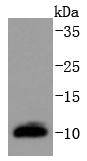
Fig1: Western blot analysis of DYNLL1 on MCF-7 cells lysates using anti-DYNLL1 antibody at 1/1,000 dilution.Application
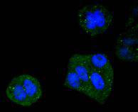
Fig2: ICC staining DYNLL1 in Hela cells (green). The nuclear counter stain is DAPI (blue). Cells were fixed in paraformaldehyde, permeabilised with 0.25% Triton X100/PBS.Application
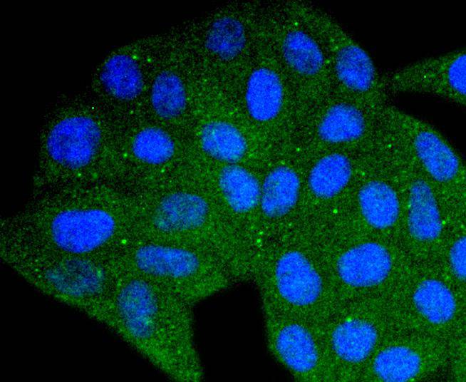
Fig3: ICC staining DYNLL1 in HepG2 cells (green). The nuclear counter stain is DAPI (blue). Cells were fixed in paraformaldehyde, permeabilised with 0.25% Triton X100/PBS.Application
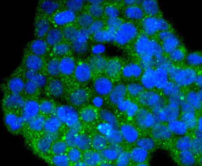
Fig4: ICC staining DYNLL1 in 293T cells (green). The nuclear counter stain is DAPI (blue). Cells were fixed in paraformaldehyde, permeabilised with 0.25% Triton X100/PBS.Application
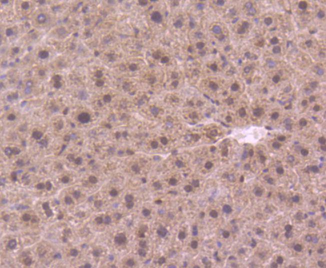
Fig5: Immunohistochemical analysis of paraffin-embedded mouse liver tissue using anti-DYNLL1 antibody. Counter stained with hematoxylin.Application
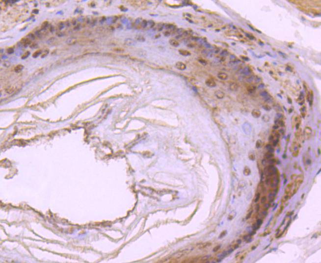
Fig6: Immunohistochemical analysis of paraffin-embedded rat esophagus tissue using anti-DYNLL1 antibody. Counter stained with hematoxylin.| Product Name | DYNLL1 [SD08-04] |
|---|---|
| Antibody Type | Primary Antibodies |
| Antigen Alias | 8 kDa dynein light chain antibody 8kDLC antibody Cytoplasmic dynein light polypeptide antibody DLC1 antibody DLC8 antibody DNCL1 antibody DNCLC1 antibody DYL1_HUMAN antibody Dynein , cytoplasmic, light chain 1 antibody Dynein light chain 1 cytoplasmic antibody Dynein light chain 1, cytoplasmic antibody Dynein light chain LC8 type 1 antibody Dynein light chain LC8-type 1 antibody Dynein, cytoplasmic, light polypeptide 1 antibody Dynein, light chain, LC8-type 1 antibody DYNLL1 antibody HDLC1 antibody LC8 antibody LC8a antibody MGC126137 antibody MGC126138 antibody MGC72986 antibody PIN antibody Protein inhibitor of neuronal nitric oxide synthase antibody Protein inhibitor of neuronal NOS antibody |
| Product description | Dyneins are multisubunit, high molecular weight ATPases that interact with microtubules to generate force by converting the chemical energy of ATP into the mechanical energy of movement. Cytoplasmic or axonemal Dynein heavy, intermediate, light and light-intermediate chains are all components of minus end-directed motors; the complex transports cellular cargos towards the central region of the cell. The highly conserved DYNLL proteins were originally identified as light chains for microtubule-based motor protein Dynein. In mammals there are two closely related isoforms expressed, DYNLL1 and DYNLL2 which share 93% sequence identity at the protein level. DYNLL1 (Dynein light chain 1) also designated, DLC8 or PIN (Protein inhibitor of neuronal nitric oxide synthase) has been identified as a protein that interacts with NOS1 resulting in NOS1 inhibition. Dimerization is required for NOS1 activity and DYNLL1 has been shown to destabilize the NOS1 dimer. Nitric oxide may be involved in several processes such as apoptosis, synaptogenesis and neuronal development; thus DYNLL1 is implicated in these processes as well. DYNLL1 is a ubiquitously expressed protein that exhibits high expression in testis and moderate expression in brain. |
| Immunogen | recombinant protein |
| Clonality | Monoclonal |
|---|---|
| Isotype | IgG |
| Host Species | Recombinant rabbit |
| Tested Applications | WBICC/IFIHCIP |
| WB:1:1,000-1:2,000 ICC:1:50-1:200 IHC:1:50-1:200 |
|
| Species Reactivity | HumanMouseRat |
| Concentration | 1mg/ml |
| Alternative Names | 8 kDa dynein light chain antibody
8kDLC antibody
Cytoplasmic dynein light polypeptide antibody
DLC1 antibody
DLC8 antibody
DNCL1 antibody
DNCLC1 antibody
DYL1_HUMAN antibody
Dynein cytoplasmic light chain 1 antibody Dynein light chain 1 cytoplasmic antibody Dynein light chain 1 cytoplasmic antibody Dynein light chain LC8 type 1 antibody Dynein light chain LC8-type 1 antibody Dynein cytoplasmic light polypeptide 1 antibody Dynein light chain LC8-type 1 antibody DYNLL1 antibody HDLC1 antibody LC8 antibody LC8a antibody MGC126137 antibody MGC126138 antibody MGC72986 antibody PIN antibody Protein inhibitor of neuronal nitric oxide synthase antibody Protein inhibitor of neuronal NOS antibody |
|---|---|
| Molecular Weight(MW) | 10 kDa |
| Cellular Localization | Cytoplasm, Nucleus, Mitochondrion. |
Application
Fig1: Western blot analysis of DYNLL1 on MCF-7 cells lysates using anti-DYNLL1 antibody at 1/1,000 dilution.
Application
Fig2: ICC staining DYNLL1 in Hela cells (green). The nuclear counter stain is DAPI (blue). Cells were fixed in paraformaldehyde, permeabilised with 0.25% Triton X100/PBS.
Application
Fig3: ICC staining DYNLL1 in HepG2 cells (green). The nuclear counter stain is DAPI (blue). Cells were fixed in paraformaldehyde, permeabilised with 0.25% Triton X100/PBS.
Application
Fig4: ICC staining DYNLL1 in 293T cells (green). The nuclear counter stain is DAPI (blue). Cells were fixed in paraformaldehyde, permeabilised with 0.25% Triton X100/PBS.
Application
Fig5: Immunohistochemical analysis of paraffin-embedded mouse liver tissue using anti-DYNLL1 antibody. Counter stained with hematoxylin.
Application
Fig6: Immunohistochemical analysis of paraffin-embedded rat esophagus tissue using anti-DYNLL1 antibody. Counter stained with hematoxylin.| Positive Control | 293T, MCF-7, Hela, HepG2, rat esophagus tissue, mouse liver tissue. |
|---|---|
| Application Notes | WB:1:1,000-1:2,000 ICC:1:50-1:200 IHC:1:50-1:200 |
| Form | Liquid |
|---|---|
| Storage Instructions | Store at +4℃ after thawing. Aliquot store at -20℃ or -80℃. Avoid repeated freeze / thaw cycles. |
| Storage Buffer | 1*TBS (pH7.4), 1%BSA, 40%Glycerol. Preservative: 0.05% Sodium Azide. |


