Application
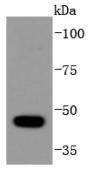
Fig1: Western blot analysis of HDAC10 on HepG2 cells lysates using anti-HDAC10 antibody at 1/1,000 dilution.Application
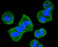
Fig2: ICC staining HDAC10 in Hela cells (green). The nuclear counter stain is DAPI (blue). Cells were fixed in paraformaldehyde, permeabilised with 0.25% Triton X100/PBS.Application
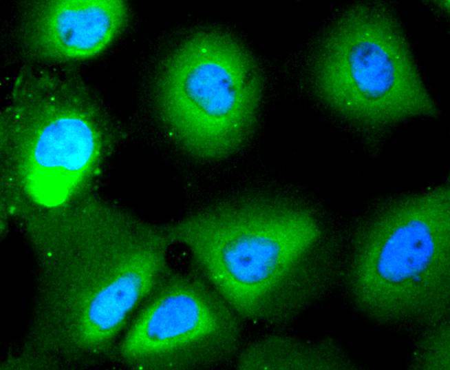
Fig3: ICC staining HDAC10 in A431 cells (green). The nuclear counter stain is DAPI (blue). Cells were fixed in paraformaldehyde, permeabilised with 0.25% Triton X100/PBS.Application
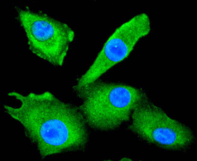
Fig4: ICC staining HDAC10 in A549 cells (green). The nuclear counter stain is DAPI (blue). Cells were fixed in paraformaldehyde, permeabilised with 0.25% Triton X100/PBS.| Product Name | HDAC10 [SD08-11] |
|---|---|
| Antibody Type | Primary Antibodies |
| Antigen Alias | DKFZP761B039 antibody HD 10 antibody HD10 antibody HDA10_HUMAN antibody HDAC 10 antibody Hdac10 antibody Histone deacetylase 10 antibody MGC149722 antibody OTTHUMP00000028555 antibody |
| Product description | Histone deacetylases (HDACs) play an important role in the modification of chromatin structure and thus in the suppression and activation of transcription and cellular differentiation. There are 11 members in the HDAC family that are divided into four classes. Class I HDACs represent homologs of the yeast histone deacetylase Rpd3, class II HDACs share strong homology with the yeast histone deacetylase Hda1, class III HDACs are closely related to the yeast Sir2 protein and class IV HDACs comprise histone deacetylase 11 (HDAC11)-related enzymes. HDAC10, also known as HD10, is a member of the class II HDACs. It contains an N-terminal Hda1p-related catalytic domain and a unique C-terminal leucine-rich domain. HDAC10 is ubiquitously expressed and can shuttle between the cytoplasm and nucleus in response to celllular signals. It is able to repress transcription and, like other class II HDAC members, its enzymatic activity is inhibited by Trichostatin A (TSA). |
| Immunogen | recombinant protein |
| Clonality | Monoclonal |
|---|---|
| Isotype | IgG |
| Host Species | Recombinant rabbit |
| Tested Applications | WBICCIP |
| WB:1:1,000 ICC:1:50-1:200 |
|
| Species Reactivity | HumanMouse |
| Concentration | 1mg/ml |
| Alternative Names | DKFZP761B039 antibody HD 10 antibody HD10 antibody HDA10_HUMAN antibody HDAC 10 antibody Hdac10 antibody Histone deacetylase 10 antibody MGC149722 antibody OTTHUMP00000028555 antibody |
|---|---|
| Molecular Weight(MW) | 43 kDa |
| Cellular Localization | Cytoplasm, Nucleus. |
Application
Fig1: Western blot analysis of HDAC10 on HepG2 cells lysates using anti-HDAC10 antibody at 1/1,000 dilution.
Application
Fig2: ICC staining HDAC10 in Hela cells (green). The nuclear counter stain is DAPI (blue). Cells were fixed in paraformaldehyde, permeabilised with 0.25% Triton X100/PBS.
Application
Fig3: ICC staining HDAC10 in A431 cells (green). The nuclear counter stain is DAPI (blue). Cells were fixed in paraformaldehyde, permeabilised with 0.25% Triton X100/PBS.
Application
Fig4: ICC staining HDAC10 in A549 cells (green). The nuclear counter stain is DAPI (blue). Cells were fixed in paraformaldehyde, permeabilised with 0.25% Triton X100/PBS.| Positive Control | A431, A549, Hela, HepG2. |
|---|---|
| Application Notes | WB:1:1,000 ICC:1:50-1:200 |
| Form | Liquid |
|---|---|
| Storage Instructions | Store at +4℃ after thawing. Aliquot store at -20℃ or -80℃. Avoid repeated freeze / thaw cycles. |
| Storage Buffer | 1*TBS (pH7.4), 1%BSA, 40%Glycerol. Preservative: 0.05% Sodium Azide. |


