Application
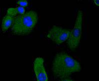
Fig1: ICC staining RON in A549 cells (green). The nuclear counter stain is DAPI (blue). Cells were fixed in paraformaldehyde, permeabilised with 0.25% Triton X100/PBS.Application
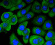
Fig2: ICC staining RON in AGS cells (green). The nuclear counter stain is DAPI (blue). Cells were fixed in paraformaldehyde, permeabilised with 0.25% Triton X100/PBS.Application
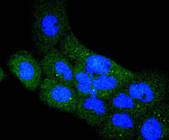
Fig3: ICC staining RON in A431 cells (green). The nuclear counter stain is DAPI (blue). Cells were fixed in paraformaldehyde, permeabilised with 0.25% Triton X100/PBS.Application
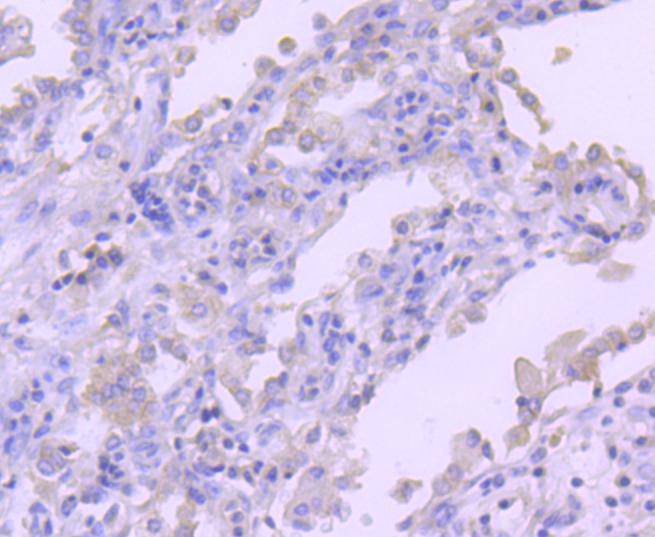
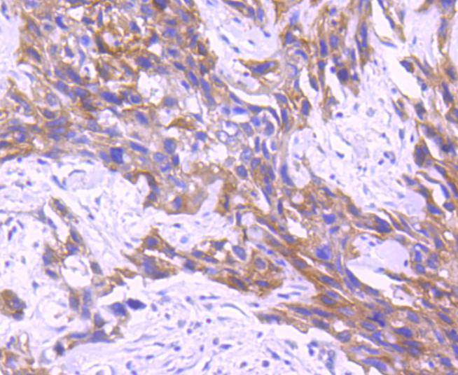
Fig4: Immunohistochemical analysis of paraffin-embedded human lung cancer tissue using anti-RON antibody. Counter stained with hematoxylin.Application
Fig5: Immunohistochemical analysis of paraffin-embedded human breast carcinoma tissue using anti-RON antibody. Counter stained with hematoxylin.Application
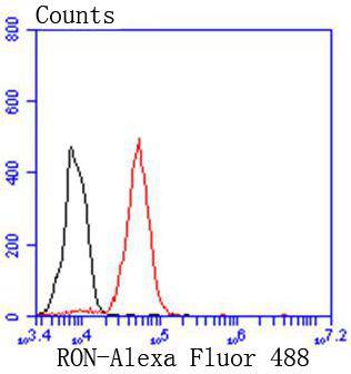
Fig6: Flow cytometric analysis of THP-1 cells with RON antibody at 1/50 dilution (red) compared with an unlabelled control (cells without incubation with primary antibody; black). Alexa Fluor 488-conjugated goat anti rabbit IgG was used as the secondary antibody| Product Name | RON [SD2006] |
|---|---|
| Antibody Type | Primary Antibodies |
| Antigen Alias | /br> Macrophage stimulating 1 receptor antibody Macrophage stimulating protein receptor alpha chain antibody MACROPHAGE STIMULATING PROTEIN RECEPTOR antibody Macrophage stimulating protein receptor beta chain antibody Macrophage-Stimulating 1 Receptor (MST1R) antibody Macrophage-stimulating protein receptor beta chain antibody MSP receptor antibody Mst1r antibody MST1R variant RON30 antibody MST1R variant RON62 antibody NPCA3 antibody p185 RON antibody p185-Ron antibody Protein-tyrosine kinase 8 antibody PTK 8 antibody ptk8 antibody PTK8 protein tyrosine kinase 8 antibody Recepteur d’origine nantais (RON) antibody RON antibody RON protein tyrosine kinase antibody RON variant E2E3 antibody RON_HUMAN antibody Soluble RON variant 1 antibody Soluble RON variant 2 antibody Soluble RON variant 3 antibody Soluble RON variant 4 antibody Stem cell derived tyrosine kinase antibody |
| Product description | Receptor protein tyrosine kinases (PTKs) have been classified into different subclasses on the basis of sequence similarity and distinct structural characteristics. The c-Met encoded receptor represents the initial member of one class of receptors characterized by a heterodimeric structure and a cysteine-rich extracellular domain. Ron, also designated macrophage-stimulating protein receptor (MSP receptor), p185-Ron, CD136 antigen or PTK8 represents a second member of this receptor class. The intracellular PTK domains of Ron and Met are highly similar (63% sequence identity) while the extracellular domains are less related (25% sequence identity) and both are rich in cysteine residues. Mature Ron receptor is comprised of a disulfide-linked heterodimer formed from an alpha chain (Ron α) and a beta chain (Ron β). Proteolytic processing results in the separation of the N-terminal Ron α and C-terminal Ron β subunits. |
| Immunogen | recombinant protein |
| Clonality | Monoclonal |
|---|---|
| Isotype | IgG |
| Host Species | Recombinant rabbit |
| Tested Applications | WBICC/IFIHCIPFC |
| WB:1:1,000 ICC:1:50-1:200 IHC:1:50-1:200 FC:1:50-1:100 |
|
| Species Reactivity | Human |
| Concentration | 1mg/ml |
| Alternative Names | /br> Macrophage stimulating 1 receptor antibody Macrophage stimulating protein receptor alpha chain antibody MACROPHAGE STIMULATING PROTEIN RECEPTOR antibody Macrophage stimulating protein receptor beta chain antibody Macrophage-Stimulating 1 Receptor (MST1R) antibody Macrophage-stimulating protein receptor beta chain antibody MSP receptor antibody Mst1r antibody MST1R variant RON30 antibody MST1R variant RON62 antibody NPCA3 antibody p185 RON antibody p185-Ron antibody Protein-tyrosine kinase 8 antibody PTK 8 antibody ptk8 antibody PTK8 protein tyrosine kinase 8 antibody Recepteur d’origine nantais (RON) antibody RON antibody RON protein tyrosine kinase antibody RON variant E2E3 antibody RON_HUMAN antibody Soluble RON variant 1 antibody Soluble RON variant 2 antibody Soluble RON variant 3 antibody Soluble RON variant 4 antibody Stem cell derived tyrosine kinase antibody |
|---|---|
| Molecular Weight(MW) | 185 kDa |
| Cellular Localization | Membrane. |
| SwissProt ID | Q04912 |
|---|
Application
Fig1: ICC staining RON in A549 cells (green). The nuclear counter stain is DAPI (blue). Cells were fixed in paraformaldehyde, permeabilised with 0.25% Triton X100/PBS.
Application
Fig2: ICC staining RON in AGS cells (green). The nuclear counter stain is DAPI (blue). Cells were fixed in paraformaldehyde, permeabilised with 0.25% Triton X100/PBS.
Application
Fig3: ICC staining RON in A431 cells (green). The nuclear counter stain is DAPI (blue). Cells were fixed in paraformaldehyde, permeabilised with 0.25% Triton X100/PBS.
Application
Fig4: Immunohistochemical analysis of paraffin-embedded human lung cancer tissue using anti-RON antibody. Counter stained with hematoxylin.
Application
Fig5: Immunohistochemical analysis of paraffin-embedded human breast carcinoma tissue using anti-RON antibody. Counter stained with hematoxylin.
Application
Fig6: Flow cytometric analysis of THP-1 cells with RON antibody at 1/50 dilution (red) compared with an unlabelled control (cells without incubation with primary antibody; black). Alexa Fluor 488-conjugated goat anti rabbit IgG was used as the secondary antibody| Positive Control | A549, AGS, A431, human lung cancer tissue, human breast carcinoma tissue. |
|---|---|
| Application Notes | WB:1:1,000 ICC:1:50-1:200 IHC:1:50-1:200 FC:1:50-1:100 |
| Form | Liquid |
|---|---|
| Storage Instructions | Store at +4℃ after thawing. Aliquot store at -20℃ or -80℃. Avoid repeated freeze / thaw cycles. |
| Storage Buffer | 1*TBS (pH7.4), 1%BSA, 40%Glycerol. Preservative: 0.05% Sodium Azide. |


