Application
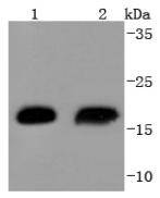
Fig1: Western blot analysis of Hsp20 on different lysates using anti-Hsp20 antibody at 1/1,000 dilution. Positive control: Lane 1: Human skeletal muscle Lane 2: Mouse skeletal muscleApplication
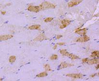
Fig2: Immunohistochemical analysis of paraffin-embedded mouse skeletal muscle tissue using anti-Hsp20 antibody. Counter stained with hematoxylin.Application
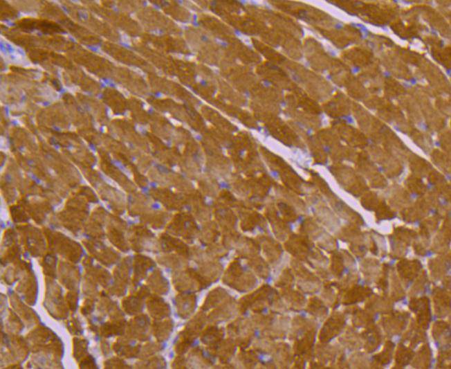
Fig3: Immunohistochemical analysis of paraffin-embedded mouse heart tissue using anti-Hsp20 antibody. Counter stained with hematoxylin.Application
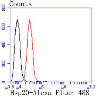
Fig4: Flow cytometric analysis of Hela cells with Hsp20 antibody at 1/50 dilution (red) compared with an unlabelled control (cells without incubation with primary antibody; black). Alexa Fluor 488-conjugated goat anti rabbit IgG was used as the secondary antibody| Product Name | Hsp20 [SD086-03] |
|---|---|
| Antibody Type | Primary Antibodies |
| Antigen Alias | epididymis luminal protein 55 antibody FLJ32389 antibody Heat shock 20 kDa like protein p20 antibody Heat shock 20 kDa-like protein p20 antibody Heat shock protein alpha crystallin related B6 antibody Heat shock protein beta 6 antibody Heat shock protein beta-6 antibody Heat shock protein, 20-KD antibody Heat-shock 27-KD protein 6 antibody HEL55 antibody Hsp20 antibody HspB6 antibody HSPB6_HUMAN antibody |
| Product description | The heat shock proteins (HSPs) comprise a group of highly conserved, abundantly expressed proteins with diverse functions, including the assembly and sequestering of multiprotein complexes, transportation of nascent poly-peptide chains across cellular membranes and regulation of protein folding. Heat shock proteins (also known as molecular chaperones) fall into six general families: HSP 90, HSP 70, HSP 60, the low molecular weight HSPs, the immunophilins and the HSP 110 family. The low molecular weight family includes HSP 10, HSP 20, HSP 27 (Heme Oxygenase 1), HSP 32 and HSP 40. HSP 20 occurs in two complex sizes, dimers and multimers. It is related to stress proteins and occurs most abundantly in skeletal muscle and heart. HSP 20 is considerably shorter at the C-terminus and less polar than other small heat shock proteins. |
| Immunogen | recombinant protein |
| Clonality | Monoclonal |
|---|---|
| Isotype | IgG |
| Host Species | Recombinant rabbit |
| Tested Applications | WBIHCFC |
| WB:1:1,000-1:2,000 IHC:1:50-1:500: FC:1:50-1:100 |
|
| Species Reactivity | HumanMouseRat |
| Concentration | 1mg/ml |
| Alternative Names | epididymis luminal protein 55 antibody
FLJ32389 antibody
Heat shock 20 kDa like protein p20 antibody
Heat shock 20 kDa-like protein p20 antibody
Heat shock protein alpha crystallin related B6 antibody
Heat shock protein beta 6 antibody
Heat shock protein beta-6 antibody
Heat shock protein 20-KD antibody Heat-shock 27-KD protein 6 antibody HEL55 antibody Hsp20 antibody HspB6 antibody HSPB6_HUMAN antibody |
|---|---|
| Molecular Weight(MW) | 17 kDa |
| Cellular Localization | Cytoplasm, Nucleus. |
Application
Fig1: Western blot analysis of Hsp20 on different lysates using anti-Hsp20 antibody at 1/1,000 dilution. Positive control: Lane 1: Human skeletal muscle Lane 2: Mouse skeletal muscle
Application
Fig2: Immunohistochemical analysis of paraffin-embedded mouse skeletal muscle tissue using anti-Hsp20 antibody. Counter stained with hematoxylin.
Application
Fig3: Immunohistochemical analysis of paraffin-embedded mouse heart tissue using anti-Hsp20 antibody. Counter stained with hematoxylin.
Application
Fig4: Flow cytometric analysis of Hela cells with Hsp20 antibody at 1/50 dilution (red) compared with an unlabelled control (cells without incubation with primary antibody; black). Alexa Fluor 488-conjugated goat anti rabbit IgG was used as the secondary antibody| Positive Control | Human skeletal muscle tissue, mouse skeletal muscle tissue, mouse heart tissue. |
|---|---|
| Application Notes | WB:1:1,000-1:2,000 IHC:1:50-1:500: FC:1:50-1:100 |
| Form | Liquid |
|---|---|
| Storage Instructions | Store at +4℃ after thawing. Aliquot store at -20℃ or -80℃. Avoid repeated freeze / thaw cycles. |
| Storage Buffer | 1*TBS (pH7.4), 1%BSA, 40%Glycerol. Preservative: 0.05% Sodium Azide. |


