WB
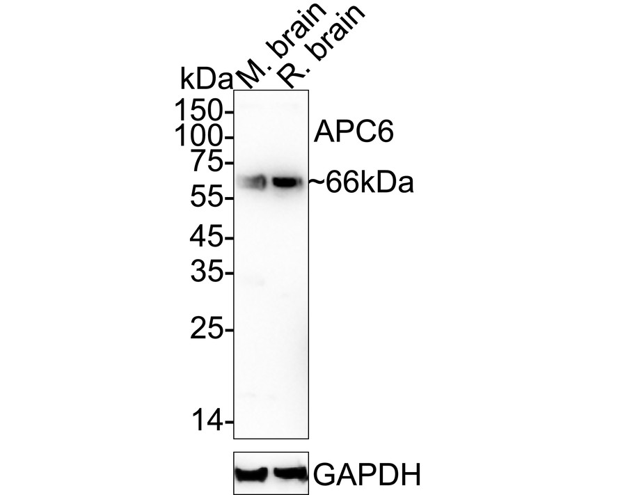
Western blot analysis of APC6 on different lysates with Rabbit anti-APC6 antibody at 1/2,000 dilution. Lane 1: Mouse brain tissue lysate, Lane 2: Rat brain tissue lysate, Lysates/proteins at 40 µg/Lane. Exposure time: 46 seconds; 4-20% SDS-PAGE gel. Proteins were transferred to a PVDF membrane and blocked with 5% NFDM/TBST for 1 hour at room temperature. The primary antibody at 1/2,000 dilution was used in 5% NFDM/TBST at 4℃ overnight. Goat Anti-Rabbit IgG - HRP Secondary Antibody at 1/50,000 dilution was used for 1 hour at room temperature.WB
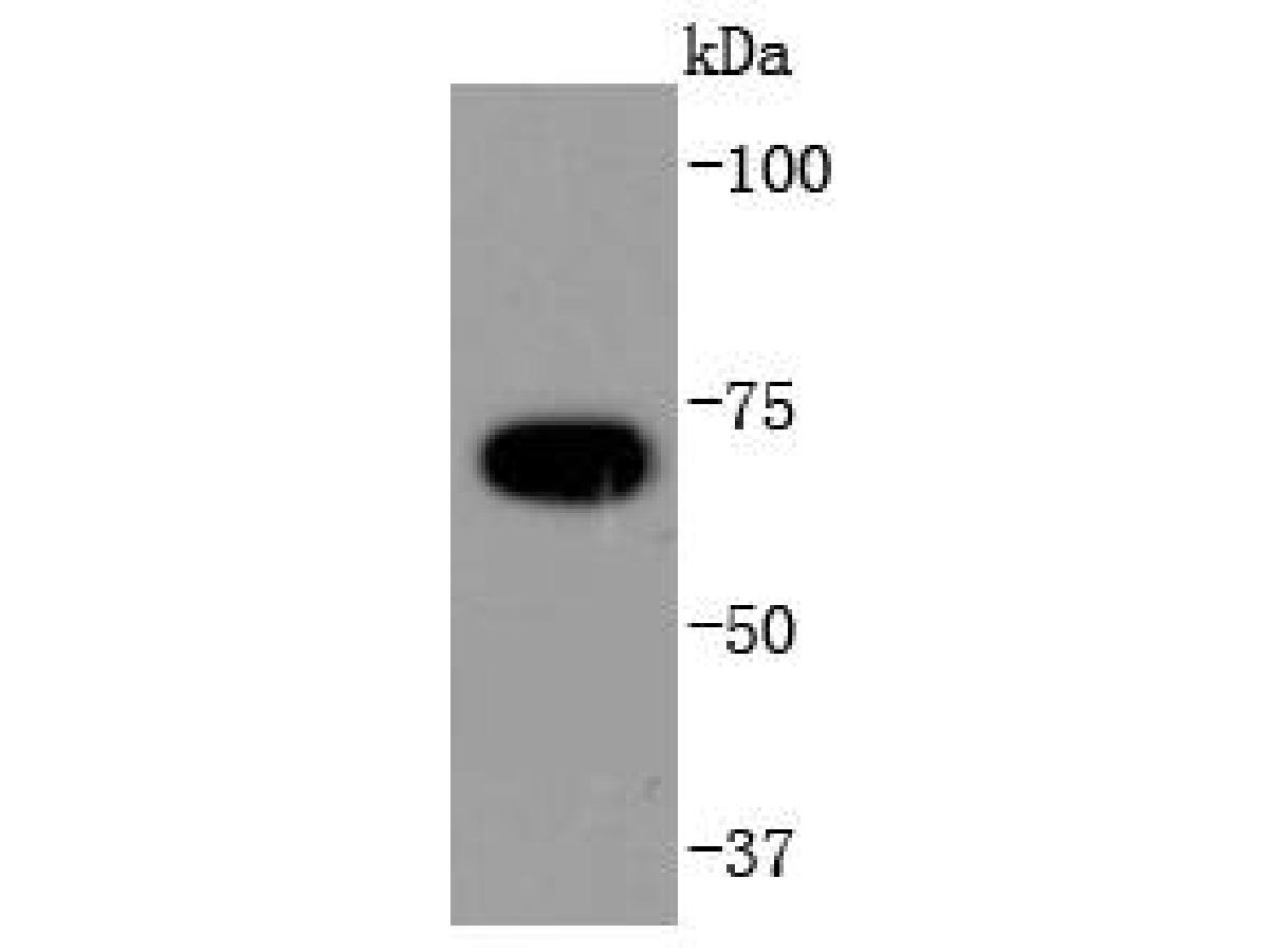
Western blot analysis of APC6 on Hela cells lysates using anti-APC6 antibody at 1/1,000 dilution.| Product Name | APC6 Recombinant Rabbit Monoclonal Antibody |
|---|---|
| Antibody Type | Primary Antibodies |
| Product description | Cell cycle events are regulated by the sequential activation and deactivation of cyclin dependent kinases (Cdks) and by the proteolysis of cyclins. The cell division cycle (Cdc) genes are required at various points in the cell cycle. Cdc25A, Cdc25B and Cdc25C protein tyrosine phosphatases function as mitotic activators by dephosphorylating Cdc2 p34 on regulatory tyrosine residues. Cdc6 is the human homolog of Saccharomyces cerevisiae Cdc6, which is involved in the initiation of DNA replication. Cdc37 appears to facilitate Cdk4/cyclin D1 complex formation and has been shown to form a stable complex with Hsp90. Cdc34, Cdc27 and Cdc16 function as ubiquitin-conjugating enzymes. Cdc34 is thought to be the structural and functional homolog of Saccharomyces cerevisiae Cdc34, which is essential for the G1 to S phase transition. Cdc16 and Cdc27 are components of the APC (anaphase-promoting complex) which ubiquitinates cyclin B, resulting in cyclin B/Cdk complex degradation. |
| Immunogen | Recombinant protein within Human APC6 aa 181-247 / 620. |
| Clonality | Monoclonal |
|---|---|
| Isotype | IgG |
| Host Species | Recombinant rabbit |
| Tested Applications | WB |
| WB:1:1000-1:2000 |
|
| Species Reactivity | HumanMouseRat |
| Concentration | 1mg/ml |
| Purification | Protein A |
| Alternative Names | ANAPC6 antibody Anaphase promoting complex subunit 6 antibody Anaphase-promoting complex subunit 6 antibody Apc 6 antibody APC6 antibody CDC 16 antibody CDC16 (cell division cycle 16 S. cerevisiae homolog) antibody Cdc16 antibody CDC16 homolog antibody CDC16 protein antibody CDC16_HUMAN antibody CDC16Hs antibody Cell division cycle 16 antibody Cell division cycle 16 homolog antibody Cell division cycle protein 16 homolog antibody CUT9 antibody Cyclosome subunit 6 antibody |
|---|---|
| Molecular Weight(MW) | 72kDa(Observed band size: 66kDa) |
| Cellular Localization | Cytoplasm. |
WB
Western blot analysis of APC6 on different lysates with Rabbit anti-APC6 antibody at 1/2,000 dilution. Lane 1: Mouse brain tissue lysate, Lane 2: Rat brain tissue lysate, Lysates/proteins at 40 µg/Lane. Exposure time: 46 seconds; 4-20% SDS-PAGE gel. Proteins were transferred to a PVDF membrane and blocked with 5% NFDM/TBST for 1 hour at room temperature. The primary antibody at 1/2,000 dilution was used in 5% NFDM/TBST at 4℃ overnight. Goat Anti-Rabbit IgG - HRP Secondary Antibody at 1/50,000 dilution was used for 1 hour at room temperature.
WB
Western blot analysis of APC6 on Hela cells lysates using anti-APC6 antibody at 1/1,000 dilution.| Application Notes | WB:1:1000-1:2000 |
|---|
| Form | Liquid |
|---|---|
| Storage Instructions | Store at +4℃ after thawing. Aliquot store at -20℃ or -80℃. Avoid repeated freeze / thaw cycles. |
| Storage Buffer | 1*TBS (pH7.4), 1%BSA, 40%Glycerol. Preservative: 0.05% Sodium Azide. |


