Application
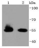
Fig1: Western blot analysis of HSPA14 on different lysates using anti-HSPA14 antibody at 1/1,000 dilution. Positive control: Lane 1: K562 Lane 2: COS-1Application
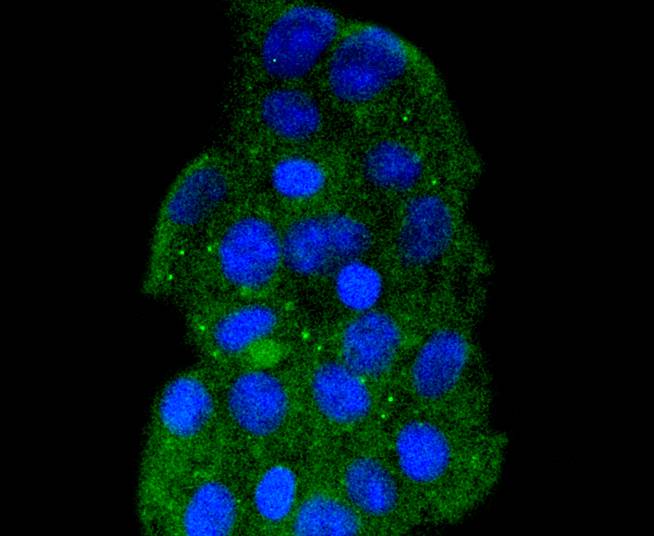
Fig2: ICC staining HSPA14 in 293 cells (green). The nuclear counter stain is DAPI (blue). Cells were fixed in paraformaldehyde, permeabilised with 0.25% Triton X100/PBS.Application
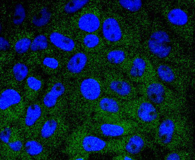
Fig3: ICC staining HSPA14 in RH-35 cells (green). The nuclear counter stain is DAPI (blue). Cells were fixed in paraformaldehyde, permeabilised with 0.25% Triton X100/PBS.Application
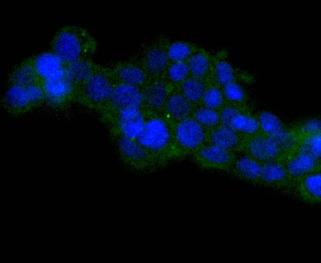
Fig4: ICC staining HSPA14 in 293T cells (green). The nuclear counter stain is DAPI (blue). Cells were fixed in paraformaldehyde, permeabilised with 0.25% Triton X100/PBS.Application
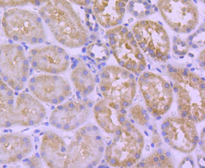
Fig5: Immunohistochemical analysis of paraffin-embedded human kidney tissue using anti-HSPA14 antibody. Counter stained with hematoxylin.Application
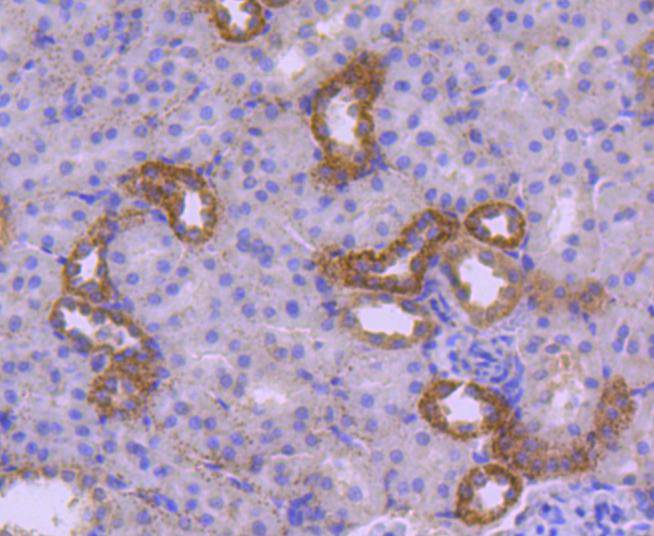
Fig6: Immunohistochemical analysis of paraffin-embedded rat kidney tissue using anti-HSPA14 antibody. Counter stained with hematoxylin.Application
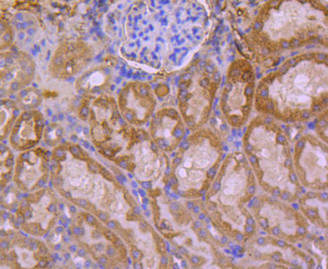
Fig7: Immunohistochemical analysis of paraffin-embedded mouse kidney tissue using anti-HSPA14 antibody. Counter stained with hematoxylin.| Product Name | HSPA14 [SD08-48] |
|---|---|
| Antibody Type | Primary Antibodies |
| Antigen Alias | Heat shock 70 kDa protein 14 antibody Heat shock 70kDa protein 14 antibody Heat shock protein HSP60 antibody Heat shock protein hsp70 related protein antibody HSP60 antibody HSP70 4 antibody HSP70 like protein 1 antibody HSP70-like protein 1 antibody HSP70L1 antibody HSP7E_HUMAN antibody HSPA 14 antibody HSPA14 antibody MGC131990 antibody |
| Product description | The heat shock proteins (HSPs) comprise a group of highly conserved, abundantly expressed proteins with diverse functions, including the assembly and sequestering of multi-protein complexes, the transportation of nascent poly-peptide chains across cellular membranes and the regulation of protein folding. HSPA14 (heat shock 70kDa protein 14), also known as HSP70-4 or HSP70L1, is a 509 amino acid novel HSP protein derived from human dendritic cells. Belonging to the heat shock protein 70 family, HSPA14 is thought to promote dendritic cell maturation. It is also suggested that HSPA14 stimulates secretion of the proinflammatory cytokines interleukin 12p70 (IL-12p70), IL-1β, TNFα, and the chemokines IP-10, MIP-1α, MIP-1β, and normal T cell expressed and secreted (RANTES). |
| Immunogen | recombinant protein |
| Clonality | Monoclonal |
|---|---|
| Isotype | IgG |
| Host Species | Recombinant rabbit |
| Tested Applications | WBICC/IFIHCIP |
| WB:1:1,000-1:2,000 ICC:1:50-1:200 IHC:1:50-1:200 |
|
| Species Reactivity | HumanMouseRat |
| Concentration | 1mg/ml |
| Alternative Names | Heat shock 70 kDa protein 14 antibody Heat shock 70kDa protein 14 antibody Heat shock protein HSP60 antibody Heat shock protein hsp70 related protein antibody HSP60 antibody HSP70 4 antibody HSP70 like protein 1 antibody HSP70-like protein 1 antibody HSP70L1 antibody HSP7E_HUMAN antibody HSPA 14 antibody HSPA14 antibody MGC131990 antibody |
|---|---|
| Molecular Weight(MW) | 55 kDa |
| Cellular Localization | Cytoplasm. |
Application
Fig1: Western blot analysis of HSPA14 on different lysates using anti-HSPA14 antibody at 1/1,000 dilution. Positive control: Lane 1: K562 Lane 2: COS-1
Application
Fig2: ICC staining HSPA14 in 293 cells (green). The nuclear counter stain is DAPI (blue). Cells were fixed in paraformaldehyde, permeabilised with 0.25% Triton X100/PBS.
Application
Fig3: ICC staining HSPA14 in RH-35 cells (green). The nuclear counter stain is DAPI (blue). Cells were fixed in paraformaldehyde, permeabilised with 0.25% Triton X100/PBS.
Application
Fig4: ICC staining HSPA14 in 293T cells (green). The nuclear counter stain is DAPI (blue). Cells were fixed in paraformaldehyde, permeabilised with 0.25% Triton X100/PBS.
Application
Fig5: Immunohistochemical analysis of paraffin-embedded human kidney tissue using anti-HSPA14 antibody. Counter stained with hematoxylin.
Application
Fig6: Immunohistochemical analysis of paraffin-embedded rat kidney tissue using anti-HSPA14 antibody. Counter stained with hematoxylin.
Application
Fig7: Immunohistochemical analysis of paraffin-embedded mouse kidney tissue using anti-HSPA14 antibody. Counter stained with hematoxylin.| Positive Control | K562, COS-1, 293, 293T, RH-35, rat kidney tissue, human kidney tissue, mouse kidney tissue. |
|---|---|
| Application Notes | WB:1:1,000-1:2,000 ICC:1:50-1:200 IHC:1:50-1:200 |
| Form | Liquid |
|---|---|
| Storage Instructions | Store at +4℃ after thawing. Aliquot store at -20℃ or -80℃. Avoid repeated freeze / thaw cycles. |
| Storage Buffer | 1*TBS (pH7.4), 1%BSA, 40%Glycerol. Preservative: 0.05% Sodium Azide. |


