Application
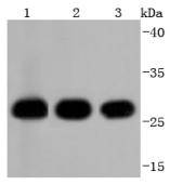
Fig1: Western blot analysis of 14-3-3 alpha+beta on different lysates using anti-14-3-3 alpha/beta antibody at 1/1,000 dilution. Positive control: Lane 1: Hela Lane 2: 293T Lane 3: HepG2Application
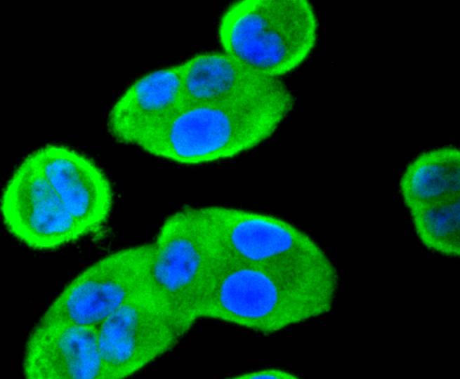
Fig2: ICC staining 14-3-3 alpha+beta in Hela cells (green). The nuclear counter stain is DAPI (blue). Cells were fixed in paraformaldehyde, permeabilised with 0.25% Triton X100/PBS.Application
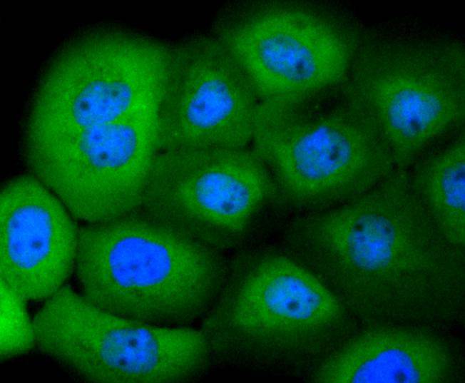
Fig3: ICC staining 14-3-3 alpha+beta in A431 cells (green). The nuclear counter stain is DAPI (blue). Cells were fixed in paraformaldehyde, permeabilised with 0.25% Triton X100/PBS.Application
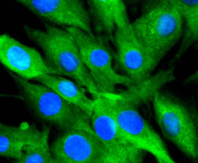
Fig4: ICC staining 14-3-3 alpha+beta in SHG-44 cells (green). The nuclear counter stain is DAPI (blue). Cells were fixed in paraformaldehyde, permeabilised with 0.25% Triton X100/PBS.Application
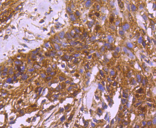
Fig5: Immunohistochemical analysis of paraffin-embedded human breast carcinoma tissue using anti-14-3-3 alpha+beta antibody. Counter stained with hematoxylin.Application
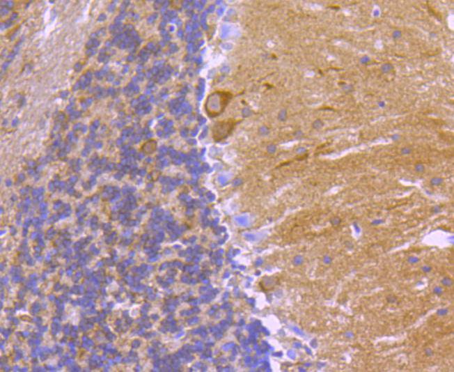
Fig6: Immunohistochemical analysis of paraffin-embedded mouse brain tissue using anti-14-3-3 alpha+beta antibody. Counter stained with hematoxylin.Application
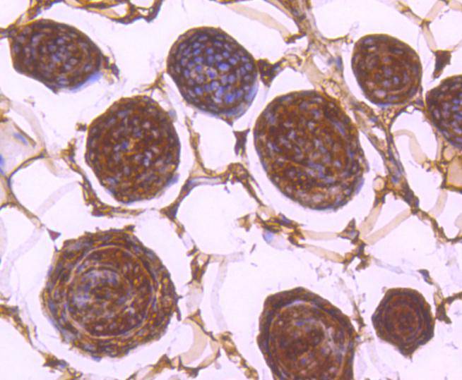
Fig7: Immunohistochemical analysis of paraffin-embedded mouse skin tissue using anti-14-3-3 alpha+beta antibody. Counter stained with hematoxylin.Application
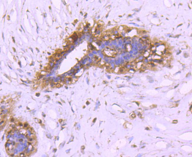
Fig8: Immunohistochemical analysis of paraffin-embedded human breast tissue using anti-14-3-3 alpha+beta antibody. Counter stained with hematoxylin.Application
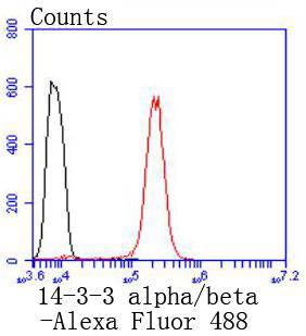
Fig9: Flow cytometric analysis of Hela cells with 14-3-3 alpha+beta antibody at 1/50 dilution (red) compared with an unlabelled control (cells without incubation with primary antibody; black). Alexa Fluor 488-conjugated goat anti rabbit IgG was used as the secondary antibody.| Product Name | 14-3-3 alpha+beta [SD0837] |
|---|---|
| Antibody Type | Primary Antibodies |
| Antigen Alias | 14 3 3 alpha antibody 14 3 3 protein beta/alpha antibody 14-3-3 protein beta/alpha antibody 1433B_HUMAN antibody Brain protein 14 3 3 beta isoform antibody GW128 antibody HS 1 antibody KCIP-1 antibody KCIP1 antibody N-terminally processed antibody Protein 1054 antibody Protein kinase C inhibitor protein 1 antibody YWHAA antibody YWHAB antibody |
| Product description | Members of the 14-3-3 family of proteins are highly conserved proteins, localized in neurons, and are axonally transported to the nerve terminals. They are also present, at lower levels, in various other eukaryotic tissues. 14-3-3 proteins appear to play important roles in a variety of signal transduction pathways, including those involved in cell cycle regulation and cell survival. Because 14-3-3 proteins bind to specific phosphoserine-containing sequences they are likely to have an important role in signaling pathways mediated by serine/threonine protein kinases. Evidence indicates 14-3-3 is required for Raf 1 kinase activity and phosphorylation among many other functions. |
| Immunogen | recombinant protein |
| Clonality | Monoclonal |
|---|---|
| Isotype | IgG |
| Host Species | Recombinant rabbit |
| Tested Applications | WBICCIHCIPFC |
| WB:1:1,000-1:5,000 ICC:1:100-1:500 IHC:1:50-1:200 FC:1:50-1:100 |
|
| Species Reactivity | HumanMouseRat |
| Concentration | 1mg/ml |
| Alternative Names | 14 3 3 alpha antibody 14 3 3 protein beta/alpha antibody 14-3-3 protein beta/alpha antibody 1433B_HUMAN antibody Brain protein 14 3 3 beta isoform antibody GW128 antibody HS 1 antibody KCIP-1 antibody KCIP1 antibody N-terminally processed antibody Protein 1054 antibody Protein kinase C inhibitor protein 1 antibody YWHAA antibody YWHAB antibody |
|---|---|
| Molecular Weight(MW) | 28 kDa |
| Cellular Localization | Cytoplasm, Melanosome. |
Application
Fig1: Western blot analysis of 14-3-3 alpha+beta on different lysates using anti-14-3-3 alpha/beta antibody at 1/1,000 dilution. Positive control: Lane 1: Hela Lane 2: 293T Lane 3: HepG2
Application
Fig2: ICC staining 14-3-3 alpha+beta in Hela cells (green). The nuclear counter stain is DAPI (blue). Cells were fixed in paraformaldehyde, permeabilised with 0.25% Triton X100/PBS.
Application
Fig3: ICC staining 14-3-3 alpha+beta in A431 cells (green). The nuclear counter stain is DAPI (blue). Cells were fixed in paraformaldehyde, permeabilised with 0.25% Triton X100/PBS.
Application
Fig4: ICC staining 14-3-3 alpha+beta in SHG-44 cells (green). The nuclear counter stain is DAPI (blue). Cells were fixed in paraformaldehyde, permeabilised with 0.25% Triton X100/PBS.
Application
Fig5: Immunohistochemical analysis of paraffin-embedded human breast carcinoma tissue using anti-14-3-3 alpha+beta antibody. Counter stained with hematoxylin.
Application
Fig6: Immunohistochemical analysis of paraffin-embedded mouse brain tissue using anti-14-3-3 alpha+beta antibody. Counter stained with hematoxylin.
Application
Fig7: Immunohistochemical analysis of paraffin-embedded mouse skin tissue using anti-14-3-3 alpha+beta antibody. Counter stained with hematoxylin.
Application
Fig8: Immunohistochemical analysis of paraffin-embedded human breast tissue using anti-14-3-3 alpha+beta antibody. Counter stained with hematoxylin.
Application
Fig9: Flow cytometric analysis of Hela cells with 14-3-3 alpha+beta antibody at 1/50 dilution (red) compared with an unlabelled control (cells without incubation with primary antibody; black). Alexa Fluor 488-conjugated goat anti rabbit IgG was used as the secondary antibody.| Positive Control | A431, SHG-44, Hela, HepG2, 293T, human breast tissue, human breast carcinoma tissue, mouse brain tissue, mouse skin tissue. |
|---|---|
| Application Notes | WB:1:1,000-1:5,000 ICC:1:100-1:500 IHC:1:50-1:200 FC:1:50-1:100 |
| Form | Liquid |
|---|---|
| Storage Instructions | Store at +4℃ after thawing. Aliquot store at -20℃ or -80℃. Avoid repeated freeze / thaw cycles. |
| Storage Buffer | 1*TBS (pH7.4), 1%BSA, 40%Glycerol. Preservative: 0.05% Sodium Azide. |


