Application
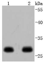
Fig1: Western blot analysis of 14-3-3 epsilon on different lysates using anti-14-3-3 epsilon antibody at 1/1,000 dilution. Positive control: Lane 1: SH-SY-5Y Lane 2: 293TApplication
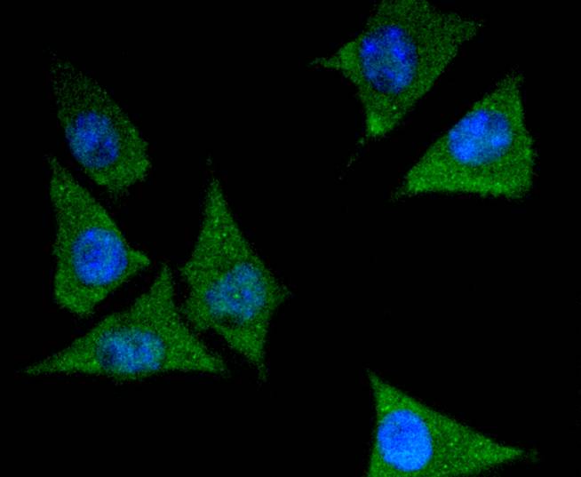
Fig2: ICC staining 14-3-3 epsilon in Hela cells (green). The nuclear counter stain is DAPI (blue). Cells were fixed in paraformaldehyde, permeabilised with 0.25% Triton X100/PBS.Application
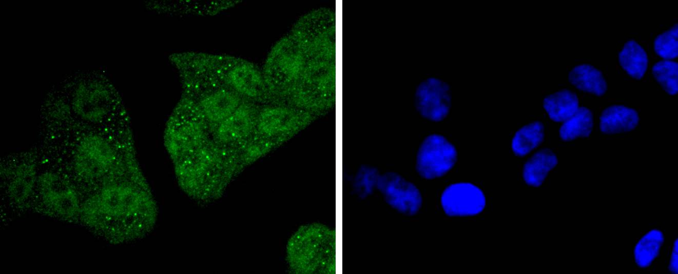
Fig3: ICC staining 14-3-3 epsilon in SH-SY-5Y cells (green). The nuclear counter stain is DAPI (blue). Cells were fixed in paraformaldehyde, permeabilised with 0.25% Triton X100/PBS.Application
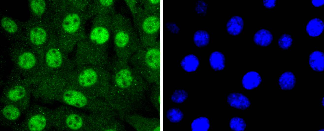
Fig4: ICC staining 14-3-3 epsilon in NIH/3T3 cells (green). The nuclear counter stain is DAPI (blue). Cells were fixed in paraformaldehyde, permeabilised with 0.25% Triton X100/PBS.Application
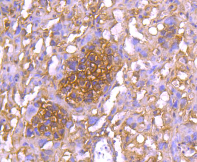
Fig5: Immunohistochemical analysis of paraffin-embedded mouse placenta tissue using anti-14-3-3 epsilon antibody. Counter stained with hematoxylin.Application
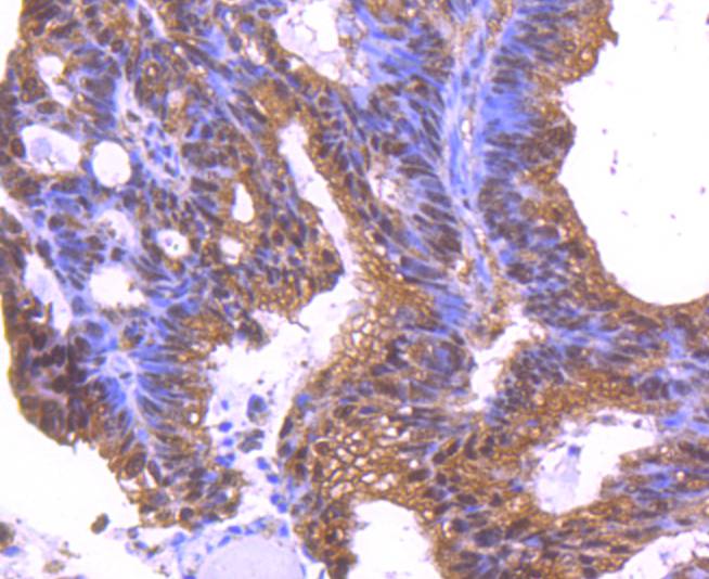
Fig6: Immunohistochemical analysis of paraffin-embedded human colon cancer tissue using anti-14-3-3 epsilon antibody. Counter stained with hematoxylin.Application
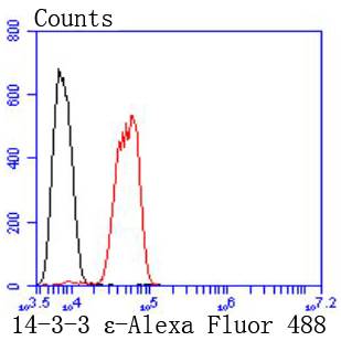
Fig7: Flow cytometric analysis of SH-SY-5Y cells with 14-3-3 epsilon antibody at 1/50 dilution (red) compared with an unlabelled control (cells without incubation with primary antibody; black). Alexa Fluor 488-conjugated goat anti rabbit IgG was used as the secondary antibody.| Product Name | 14-3-3 epsilon [JJ08-40] |
|---|---|
| Antibody Type | Primary Antibodies |
| Antigen Alias | 14 3 3 E antibody 14 3 3 epsilon antibody 14 3 3E antibody 14-3-3 protein epsilon antibody 14-3-3E antibody 1433E_HUMAN antibody Epididymis luminal protein 2 antibody FLJ45465 antibody FLJ53559 antibody HEL2 antibody KCIP 1 antibody KCIP1 antibody MDCR antibody MDS antibody Mitochondrial import stimulation factor L subunit antibody Protein kinase C inhibitor protein1 antibody Tyrosine 3 monooxygenase/tryptophan 5 monooxygenase activation protein, epsilon antibody Tyrosine 3 monooxygenase/tryptophan 5 monooxygenase activation protein, epsilon polypeptide antibody Tyrosine 3/tryptophan 5 monooxygenase activation protein epsilon polypeptide antibody YWHAE antibody |
| Product description | The 14-3-3 family of proteins plays a key regulatory role in signal transduction, checkpoint control, apoptotic and nutrient-sensing pathways. 14-3-3 proteins are highly conserved and ubiquitously expressed. There are at least seven isoforms, β, γ, ε, σ, ζ, τ and η that have been identified in mammals. The 14-3-3 epsilon, a subtype of the 14-3-3 family of proteins, was thought to be brain and neuron-specific. It has been shown to interact with CDC25 phosphatases, RAF1 and IRS1 proteins, suggesting its role in diverse biochemical activities related to signal transduction, such as cell division and regulation of insulin sensitivity. It has also been implicated in the pathogenesis of small cell lung cancer. |
| Immunogen | recombinant protein |
| Clonality | Monoclonal |
|---|---|
| Isotype | IgG |
| Host Species | Recombinant rabbit |
| Tested Applications | WBICC/IFIHCFC |
| WB:1:1,000-1:2,000 ICC:1:50-1:200 IHC:1:50-1:200 FC:1:10-1:100 |
|
| Species Reactivity | HumanMouseRat |
| Concentration | 1mg/ml |
| Alternative Names | 14 3 3 E antibody
14 3 3 epsilon antibody
14 3 3E antibody
14-3-3 protein epsilon antibody
14-3-3E antibody
1433E_HUMAN antibody
Epididymis luminal protein 2 antibody
FLJ45465 antibody
FLJ53559 antibody
HEL2 antibody
KCIP 1 antibody
KCIP1 antibody
MDCR antibody
MDS antibody
Mitochondrial import stimulation factor L subunit antibody
Protein kinase C inhibitor protein1 antibody
Tyrosine 3 monooxygenase/tryptophan 5 monooxygenase activation protein epsilon antibody Tyrosine 3 monooxygenase/tryptophan 5 monooxygenase activation protein epsilon polypeptide antibody Tyrosine 3/tryptophan 5 monooxygenase activation protein epsilon polypeptide antibody YWHAE antibody |
|---|---|
| Molecular Weight(MW) | 29 kDa |
| Cellular Localization | Cytoplasm, Melanosome. |
Application
Fig1: Western blot analysis of 14-3-3 epsilon on different lysates using anti-14-3-3 epsilon antibody at 1/1,000 dilution. Positive control: Lane 1: SH-SY-5Y Lane 2: 293T
Application
Fig2: ICC staining 14-3-3 epsilon in Hela cells (green). The nuclear counter stain is DAPI (blue). Cells were fixed in paraformaldehyde, permeabilised with 0.25% Triton X100/PBS.
Application
Fig3: ICC staining 14-3-3 epsilon in SH-SY-5Y cells (green). The nuclear counter stain is DAPI (blue). Cells were fixed in paraformaldehyde, permeabilised with 0.25% Triton X100/PBS.
Application
Fig4: ICC staining 14-3-3 epsilon in NIH/3T3 cells (green). The nuclear counter stain is DAPI (blue). Cells were fixed in paraformaldehyde, permeabilised with 0.25% Triton X100/PBS.
Application
Fig5: Immunohistochemical analysis of paraffin-embedded mouse placenta tissue using anti-14-3-3 epsilon antibody. Counter stained with hematoxylin.
Application
Fig6: Immunohistochemical analysis of paraffin-embedded human colon cancer tissue using anti-14-3-3 epsilon antibody. Counter stained with hematoxylin.
Application
Fig7: Flow cytometric analysis of SH-SY-5Y cells with 14-3-3 epsilon antibody at 1/50 dilution (red) compared with an unlabelled control (cells without incubation with primary antibody; black). Alexa Fluor 488-conjugated goat anti rabbit IgG was used as the secondary antibody.| Positive Control | SH-SY-5Y, NIH/3T3, Hela, 293T, human colon cancer tissue, mouse placenta tissue. |
|---|---|
| Application Notes | WB:1:1,000-1:2,000 ICC:1:50-1:200 IHC:1:50-1:200 FC:1:10-1:100 |
| Form | Liquid |
|---|---|
| Storage Instructions | Store at +4℃ after thawing. Aliquot store at -20℃ or -80℃. Avoid repeated freeze / thaw cycles. |
| Storage Buffer | 1*TBS (pH7.4), 1%BSA, 40%Glycerol. Preservative: 0.05% Sodium Azide. |


