Application
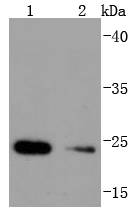
Fig1: Western blot analysis of Hsp22 on different lysates using anti-Hsp22 antibody at 1/1,000 dilution. Positive control: Lane 1: HepG2 Lane 2: Mouse skeletal muscleApplication
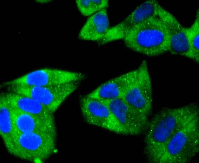
Fig2: ICC staining Hsp22 in Hela cells (green). The nuclear counter stain is DAPI (blue). Cells were fixed in paraformaldehyde, permeabilised with 0.25% Triton X100/PBS.Application
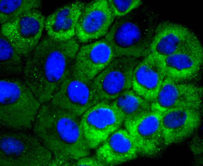
Fig3: ICC staining Hsp22 in A431 cells (green). The nuclear counter stain is DAPI (blue). Cells were fixed in paraformaldehyde, permeabilised with 0.25% Triton X100/PBS.Application
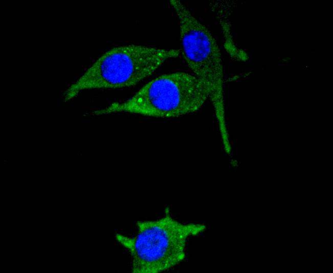
Fig4: ICC staining Hsp22 in SH-SY-5Y cells (green). The nuclear counter stain is DAPI (blue). Cells were fixed in paraformaldehyde, permeabilised with 0.25% Triton X100/PBS.Application
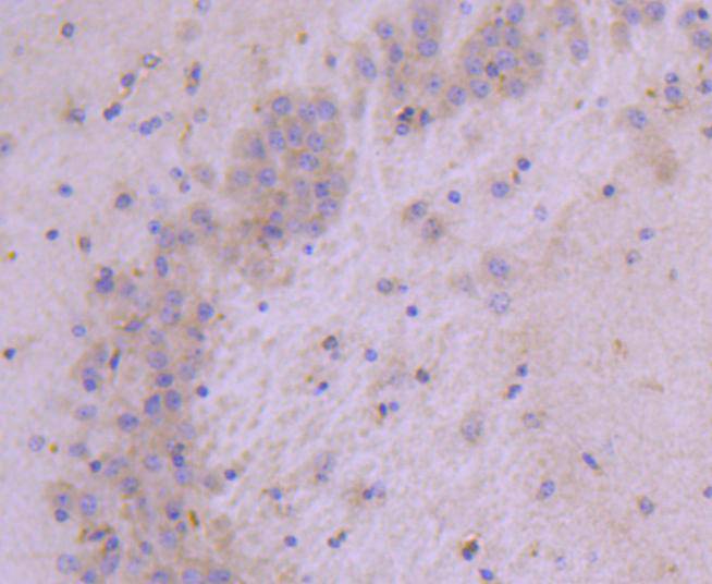
Fig5: Immunohistochemical analysis of paraffin-embedded mouse brain tissue using anti-Hsp22 antibody. Counter stained with hematoxylin.Application
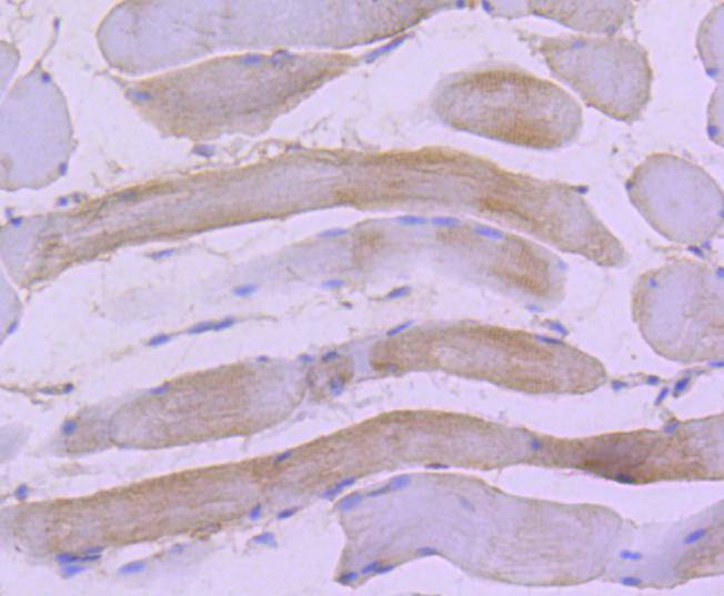
Fig6: Immunohistochemical analysis of paraffin-embedded rat skeletal muscle tissue using anti-Hsp22 antibody. Counter stained with hematoxylin.| Product Name | Hsp22 [JJ08-53] |
|---|---|
| Antibody Type | Primary Antibodies |
| Antigen Alias | Alpha crystallin C chain antibody Alpha-crystallin C chain antibody Charcot Marie Tooth disease axonal type 2L antibody Charcot Marie Tooth disease spinal antibody CMT2L antibody CRYAC antibody DHMN 2 antibody DHMN2 antibody E2 induced gene 1 protein antibody E2-induced gene 1 protein antibody E2IG1 antibody H11 antibody Heat shock 22kDa protein 8 antibody Heat shock 27kDa protein 8 antibody Heat shock protein 22 antibody Heat shock protein beta 8 antibody Heat shock protein beta-8 antibody Hereditary motor neuropathy distal antibody HMN 2 antibody HMN2 antibody HMN2A antibody HSB8 antibody HSPB 8 antibody HspB8 antibody HSPB8_HUMAN antibody OTTHUMP00000239768 antibody Protein kinase H11 antibody Small stress protein like protein HSP22 antibody Small stress protein-like protein HSP22 antibody Spinal muscular atrophy distal adult autosomal dominant antibody |
| Product description | Crystallins are the major proteins expressed in the vertebrate eye lens, where they maintain the transparency and refractive index of the lens. Crystallins are divided into a, b and g families; b and g-crystallins compose a superfamily. Crystallins usually contain seven distinctive protein regions, including four homologous motifs, a connecting peptide, and N- and C-terminal extensions. a-crystallins consist of three gene products, aA, aB and aC-crystallin, which are members of the small heat shock protein family (HSP20). They are induced by heat shock, and act as molecular chaperones by holding denatured proteins in large soluble aggregates. However, unlike other molecular chaperones, a-crystallins do not renature these proteins. Research indicates that binding occurs between membranes and aC-crystallin. The binding site appears to be at the polar-apolar interface in membrane protein (MIP26) and aC-crystallin; the lipid bilayer becomes less mobile with aC-crystallin binding. |
| Immunogen | recombinant protein |
| Clonality | Monoclonal |
|---|---|
| Isotype | IgG |
| Host Species | Recombinant rabbit |
| Tested Applications | WBICC/IFIHC |
| WB:1:1,000-1:2,000 ICC:1:100-1:500 IHC:1:50-1:200 |
|
| Species Reactivity | HumanMouseRat |
| Concentration | 1mg/ml |
| Alternative Names | Alpha crystallin C chain antibody Alpha-crystallin C chain antibody Charcot Marie Tooth disease axonal type 2L antibody Charcot Marie Tooth disease spinal antibody CMT2L antibody CRYAC antibody DHMN 2 antibody DHMN2 antibody E2 induced gene 1 protein antibody E2-induced gene 1 protein antibody E2IG1 antibody H11 antibody Heat shock 22kDa protein 8 antibody Heat shock 27kDa protein 8 antibody Heat shock protein 22 antibody Heat shock protein beta 8 antibody Heat shock protein beta-8 antibody Hereditary motor neuropathy distal antibody HMN 2 antibody HMN2 antibody HMN2A antibody HSB8 antibody HSPB 8 antibody HspB8 antibody HSPB8_HUMAN antibody OTTHUMP00000239768 antibody Protein kinase H11 antibody Small stress protein like protein HSP22 antibody Small stress protein-like protein HSP22 antibody Spinal muscular atrophy distal adult autosomal dominant antibody |
|---|---|
| Molecular Weight(MW) | 22 kDa |
| Cellular Localization | Cytoplasm, Nucleus. |
Application
Fig1: Western blot analysis of Hsp22 on different lysates using anti-Hsp22 antibody at 1/1,000 dilution. Positive control: Lane 1: HepG2 Lane 2: Mouse skeletal muscle
Application
Fig2: ICC staining Hsp22 in Hela cells (green). The nuclear counter stain is DAPI (blue). Cells were fixed in paraformaldehyde, permeabilised with 0.25% Triton X100/PBS.
Application
Fig3: ICC staining Hsp22 in A431 cells (green). The nuclear counter stain is DAPI (blue). Cells were fixed in paraformaldehyde, permeabilised with 0.25% Triton X100/PBS.
Application
Fig4: ICC staining Hsp22 in SH-SY-5Y cells (green). The nuclear counter stain is DAPI (blue). Cells were fixed in paraformaldehyde, permeabilised with 0.25% Triton X100/PBS.
Application
Fig5: Immunohistochemical analysis of paraffin-embedded mouse brain tissue using anti-Hsp22 antibody. Counter stained with hematoxylin.
Application
Fig6: Immunohistochemical analysis of paraffin-embedded rat skeletal muscle tissue using anti-Hsp22 antibody. Counter stained with hematoxylin.| Positive Control | A431, SH-SY-5Y, Hela, HepG2, rat skeletal muscle tissue, mouse skeletal muscle tissue, mouse brain tissue. |
|---|---|
| Application Notes | WB:1:1,000-1:2,000 ICC:1:100-1:500 IHC:1:50-1:200 |
| Form | Liquid |
|---|---|
| Storage Instructions | Store at +4℃ after thawing. Aliquot store at -20℃ or -80℃. Avoid repeated freeze / thaw cycles. |
| Storage Buffer | 1*TBS (pH7.4), 1%BSA, 40%Glycerol. Preservative: 0.05% Sodium Azide. |


