Application
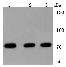
Fig1: Western blot analysis of HSPA2 on different lysates using anti-HSPA2 antibody at 1/1,000 dilution. Positive control: Lane 1: MCF-7 Lane 2: Hela Lane 3: A431Application
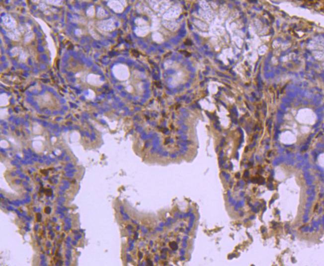
Fig2: Immunohistochemical analysis of paraffin-embedded mouse colon tissue using anti-HSPA2 antibody. Counter stained with hematoxylin.| Product Name | HSPA2 [JJ08-77] |
|---|---|
| Antibody Type | Primary Antibodies |
| Antigen Alias | 70kDa antibody Hcp70.2 antibody Heat shock 70 kDa protein 2 antibody Heat shock protein 2 antibody Heat shock protein 70.2 antibody Heat shock related 70 kDa protein 2 antibody Heat shock-related 70 kDa protein 2 antibody Heat-shock protein, 70-KD, 2 antibody Heat-shock protein, 70-KD, 3 antibody HSP70 2 antibody HSP70 3 antibody Hsp70-2 antibody HSP70-3 antibody HSP70.2 antibody HSP70A2 antibody HSP72 antibody HSP72_HUMAN antibody HSPA2 antibody Hspt70 antibody Hst70 antibody MGC58299 antibody MGC7795 antibody MGC93458 antibody OTTHUMP00000180664 antibody Testis-specific heat shock protein-related antibody |
| Product description | HSPA2(heat shock 70kDa protein 2) also known as HEAT-SHOCK PROTEIN, 70-KD, 2, HSP70-2, HEAT-SHOCK PROTEIN, 70-KD, 3 or HSP70-3. Analysis of the sequence indicated that HSPA2 is the human homolog of the murine Hsp70-2 gene, with 91.7% identity in the nucleotide coding sequence and 98.2% in the corresponding amino acid sequence.HSPA2 has less amino acid homology to the other members of the human HSP70 gene family. HSPA2 is constitutively expressed in most tissues, with very high levels in testis and skeletal muscle. The HSPA2 gene is located on chromosome 14q22-q24. Immunohistochemical analysis detected weak expression of HSPA2 in spermatocytes and stronger expression in spermatids and in the tail of mature sperm. HSPA2 may be critical to sperm maturation through its role as a protein chaperone. |
| Immunogen | recombinant protein |
| Clonality | Monoclonal |
|---|---|
| Isotype | IgG |
| Host Species | Recombinant rabbit |
| Tested Applications | WBIHC |
| WB:1:1,000-1:2,000 IHC:1:50-1:200 |
|
| Species Reactivity | HumanMouseRat |
| Concentration | 1mg/ml |
| Alternative Names | 70kDa antibody
Hcp70.2 antibody
Heat shock 70 kDa protein 2 antibody
Heat shock protein 2 antibody
Heat shock protein 70.2 antibody
Heat shock related 70 kDa protein 2 antibody
Heat shock-related 70 kDa protein 2 antibody
Heat-shock protein 70-KD 2 antibody Heat-shock protein 70-KD 3 antibody HSP70 2 antibody HSP70 3 antibody Hsp70-2 antibody HSP70-3 antibody HSP70.2 antibody HSP70A2 antibody HSP72 antibody HSP72_HUMAN antibody HSPA2 antibody Hspt70 antibody Hst70 antibody MGC58299 antibody MGC7795 antibody MGC93458 antibody OTTHUMP00000180664 antibody Testis-specific heat shock protein-related antibody |
|---|---|
| Molecular Weight(MW) | 70 kDa |
| Cellular Localization | Cytoplasm. |
Application
Fig1: Western blot analysis of HSPA2 on different lysates using anti-HSPA2 antibody at 1/1,000 dilution. Positive control: Lane 1: MCF-7 Lane 2: Hela Lane 3: A431
Application
Fig2: Immunohistochemical analysis of paraffin-embedded mouse colon tissue using anti-HSPA2 antibody. Counter stained with hematoxylin.| Positive Control | A431, MCF-7, Hela, mouse colon tissue. |
|---|---|
| Application Notes | WB:1:1,000-1:2,000 IHC:1:50-1:200 |
| Form | Liquid |
|---|---|
| Storage Instructions | Store at +4℃ after thawing. Aliquot store at -20℃ or -80℃. Avoid repeated freeze / thaw cycles. |
| Storage Buffer | 1*TBS (pH7.4), 1%BSA, 40%Glycerol. Preservative: 0.05% Sodium Azide. |


