WB
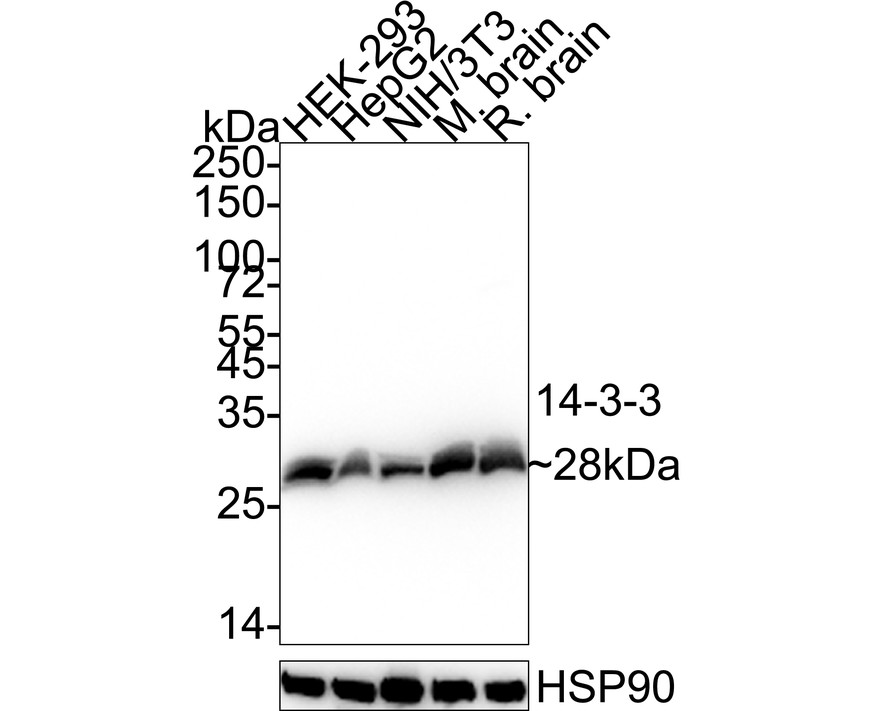
Western blot analysis of 14-3-3 on different lysates with Rabbit anti-14-3-3 antibody at 1/1,000 dilution. Lane 1: HEK-293 cell lysate (20 µg/Lane), Lane 2: HepG2 cell lysate (20 µg/Lane), Lane 3: NIH/3T3 cell lysate (20 µg/Lane), Lane 4: Mouse brain tissue lysate (40 µg/Lane), Lane 5: Rat brain tissue lysate (40 µg/Lane), Exposure time: 2 seconds; 4-20% SDS-PAGE gel. Proteins were transferred to a PVDF membrane and blocked with 5% NFDM/TBST for 1 hour at room temperature. The primary antibody at 1/1,000 dilution was used in 5% NFDM/TBST at 4℃ overnight. Goat Anti-Rabbit IgG - HRP Secondary Antibody at 1/50,000 dilution was used for 1 hour at room temperature.IHC
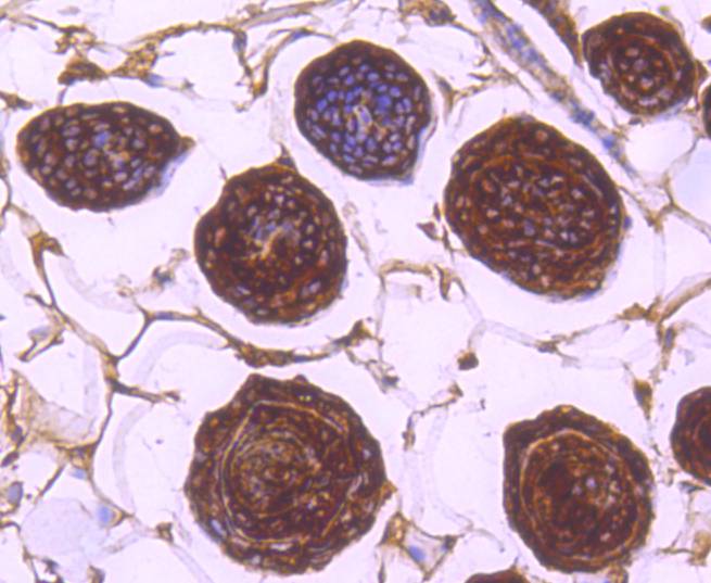
Immunohistochemical analysis of paraffin-embedded mouse skin tissue using anti-14-3-3 antibody. Counter stained with hematoxylin.| Product Name | 14-3-3 Recombinant Rabbit Monoclonal Antibody |
|---|---|
| Antibody Type | Primary Antibodies |
| Product description | The 14-3-3 family of proteins are important regulatory molecules ubiquitiously expressed in all eukarryotic cells which bind to numerous signaling proteins in various pathways driving critical cellular pathways of apoptosis, differentiation and cell cycle. 14-3-3 theta specifically has also been shown to play an important regulatory role in the TLR2 signaling pathways as a negative regulator of TLR2 ligand Pam3CySk4 induced NF-kB activation. 14-3-3 theta has previously been shown to interact with TLR4 ligand and MyD88 dependent phosphorylated PkC epsilon. 14-3-3 theta in the TLR4 signaling pathway is a positive regulator controlling release of IRF3 indcuded pro-inflammatory cytokines RANTES and IP-10. Currently identified by mass spec as part of the TLR2 signaling complex and taken along with TLR4 data, a 14-3-3 theta antibody can be used to examine the different regulatory functions of 14-3-3 theta for different TLRs through its interaction with common or unique TLR signaling adaptor molecules in addition to MyD88 or PkC epsilon such as TRAM or TRIF allowing further clarification of TLR specific pathway regulation. |
| Immunogen | Recombinant protein within Human 14-3-3 aa 1-126 / 246. |
| Clonality | Monoclonal |
|---|---|
| Isotype | IgG |
| Host Species | Recombinant rabbit |
| Tested Applications | IHCWB |
| WB:1:1000-1:5000 IHC:1:50-1:200 |
|
| Species Reactivity | HumanMouseRat |
| Concentration | 1mg/ml |
| Purification | Protein A |
| Molecular Weight(MW) | 28 kDa |
|---|---|
| Cellular Localization | Cytoplasm, Melanosome, Vacuole membrane. |
WB
Western blot analysis of 14-3-3 on different lysates with Rabbit anti-14-3-3 antibody at 1/1,000 dilution. Lane 1: HEK-293 cell lysate (20 µg/Lane), Lane 2: HepG2 cell lysate (20 µg/Lane), Lane 3: NIH/3T3 cell lysate (20 µg/Lane), Lane 4: Mouse brain tissue lysate (40 µg/Lane), Lane 5: Rat brain tissue lysate (40 µg/Lane), Exposure time: 2 seconds; 4-20% SDS-PAGE gel. Proteins were transferred to a PVDF membrane and blocked with 5% NFDM/TBST for 1 hour at room temperature. The primary antibody at 1/1,000 dilution was used in 5% NFDM/TBST at 4℃ overnight. Goat Anti-Rabbit IgG - HRP Secondary Antibody at 1/50,000 dilution was used for 1 hour at room temperature.
IHC
Immunohistochemical analysis of paraffin-embedded mouse skin tissue using anti-14-3-3 antibody. Counter stained with hematoxylin.| Application Notes | WB:1:1000-1:5000 IHC:1:50-1:200 |
|---|
| Form | Liquid |
|---|---|
| Storage Instructions | Store at +4℃ after thawing. Aliquot store at -20℃ or -80℃. Avoid repeated freeze / thaw cycles. |
| Storage Buffer | 1*TBS (pH7.4), 1%BSA, 40%Glycerol. Preservative: 0.05% Sodium Azide. |


