WB
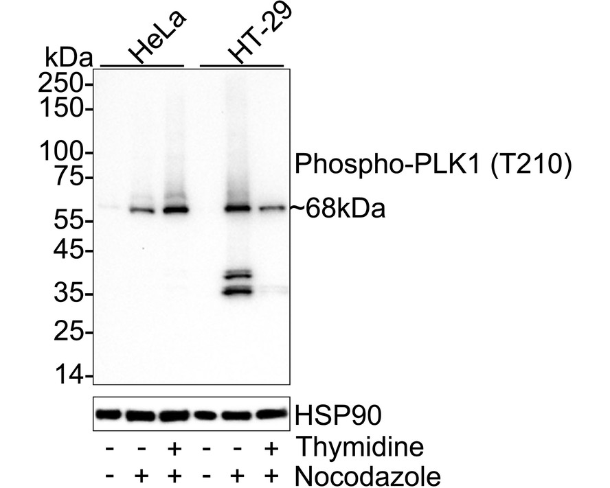
Western blot analysis of Phospho-PLK1 (T210) on different lysates with Rabbit anti-Phospho-PLK1 (T210) antibody at 1/1,000 dilution. Lane 1: HeLa cell lysate, Lane 2: HeLa treated with 100nM Nocodazole for 24 hours cell lysate, Lane 3: HeLa treated with 2mM thymidine for 16 hours then treated with 100nM Nocodazole for 24 hours cell lysate, Lane 4: HT-29 cell lysate, Lane 5: HT-29 treated with 100nM Nocodazole for 24 hours cell lysate, Lane 6: HT-29 treated with 2mM thymidine for 16 hours then treated with 100nM Nocodazole for 24 hours cell lysate, Lysates/proteins at 20 µg/Lane. Exposure time: 3 minutes; 4-20% SDS-PAGE gel. Proteins were transferred to a PVDF membrane and blocked with 5% NFDM/TBST for 1 hour at room temperature. The primary antibody at 1/1,000 dilution was used in primary antibody dilution at 4℃ overnight. Goat Anti-Rabbit IgG - HRP Secondary Antibody at 1/50,000 dilution was used for 1 hour at room temperature.IHC
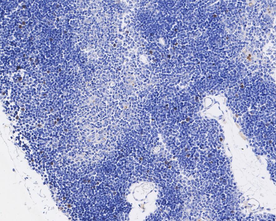
Immunohistochemical analysis of paraffin-embedded mouse thymus tissue with Rabbit anti-Phospho-PLK1 (T210) antibody at 1/200 dilution. The section was pre-treated using heat mediated antigen retrieval with Tris-EDTA buffer (pH 9.0) for 20 minutes. The tissues were blocked in 1% BSA for 20 minutes at room temperature, washed with ddH2O and PBS, and then probed with the primary antibody at 1/200 dilution for 1 hour at room temperature. The detection was performed using an HRP conjugated compact polymer system. DAB was used as the chromogen. Tissues were counterstained with hematoxylin and mounted with DPX.FC
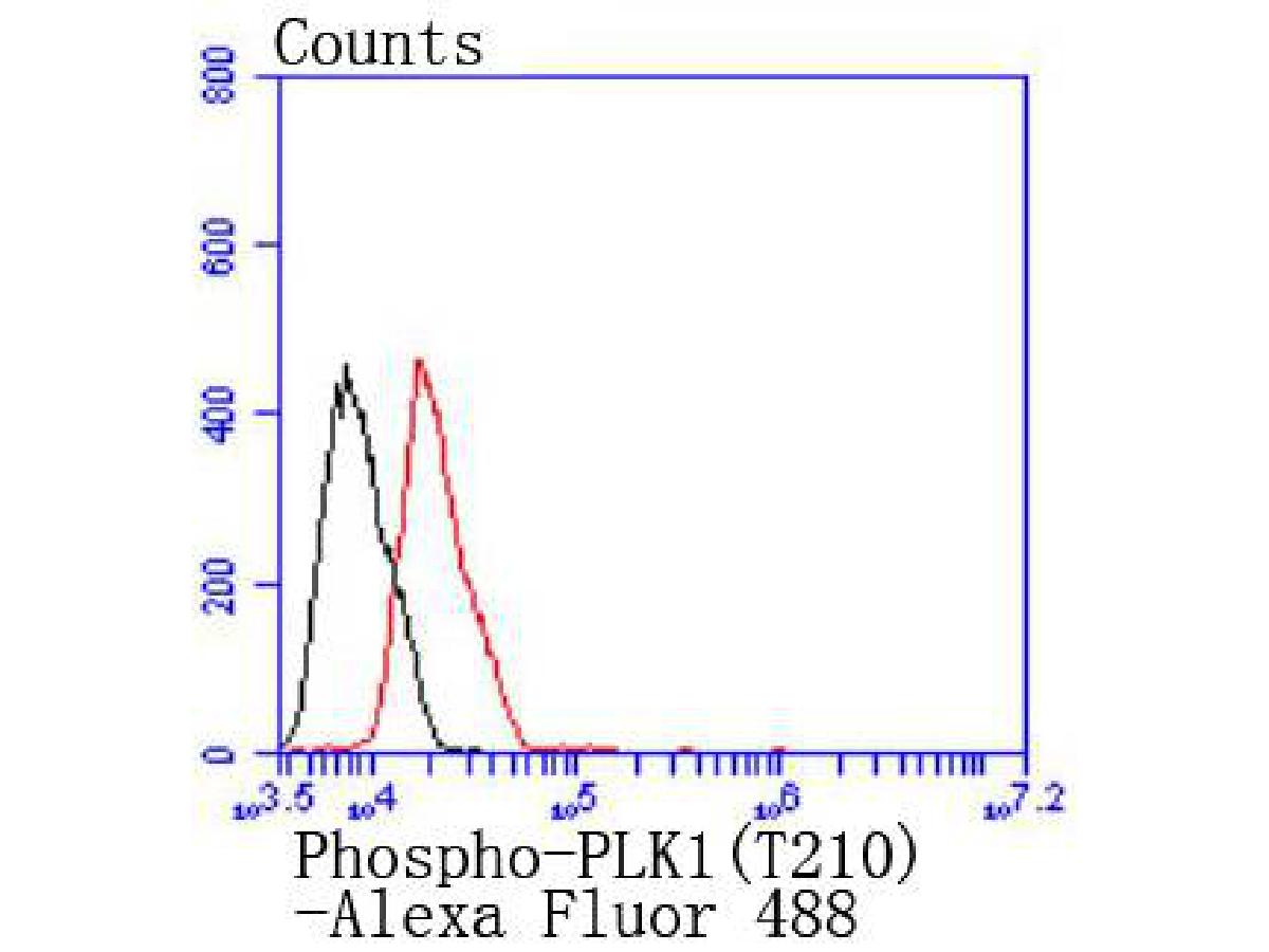
Flow cytometric analysis of Phospho-PLK1 (T210) was done on MCF-7 cells. The cells were fixed, permeabilized and stained with the primary antibody (1/50) (red). After incubation of the primary antibody at room temperature for an hour, the cells were stained with a Alexa Fluor 488-conjugated Goat anti-Rabbit IgG Secondary antibody at 1/1000 dilution for 30 minutes.Unlabelled sample was used as a control (cells without incubation with primary antibody; black).| Product Name | Phospho-PLK1 (T210) Recombinant Rabbit Monoclonal Antibody |
|---|---|
| Antibody Type | Primary Antibodies |
| Product description | Plk (for polo-like kinase) encodes a serine/threonine kinase that is closely related to polo and CDC5, genes that are required for passage through mitosis in Drosophila and Saccharomyces, respectively. Polo and CDC5 both code for proteins that are involved in regulating the function of the mitotic spindle. Plk protein accumulates in the cell during the S and G2 phases of the cell cycle and both protein content and catalytic activity peak at the onset of mitosis, followed by a rapid reduction after mitosis. Plk expression is detectable in mitotically active tissues such as colon and placenta, as well as in tumors of various origins. It has also been suggested that Plk may serve as a marker of cell proliferation. The phosphorylation of mouse, rat and human Plk on Thr 210 enhances Plk catalytic activity. |
| Immunogen | Synthetic phospho-peptide corresponding to residues surrounding Thr210 of Human PLK1 aa 181-230 / 603. |
| Modification | p-T210 |
| Clonality | Monoclonal |
|---|---|
| Isotype | IgG |
| Host Species | Recombinant rabbit |
| Tested Applications | FCIHCWB |
| WB:1:1000 IHC:1:200 ICC/IF:1:10-1:50 |
|
| Species Reactivity | HumanMouse |
| Concentration | 1mg/ml |
| Purification | Protein A |
| Alternative Names | Cell cycle regulated protein kinase antibody PLK 1 antibody PLK antibody PLK-1 antibody plk1 antibody PLK1_HUMAN antibody Polo like kinase 1 antibody Polo-like kinase 1 antibody Serine/threonine protein kinase 13 antibody Serine/threonine protein kinase PLK1 antibody Serine/threonine-protein kinase 13 antibody Serine/threonine-protein kinase PLK1 antibody STPK 13 antibody STPK13 antibody |
|---|---|
| Molecular Weight(MW) | 68 kDa |
| Cellular Localization | Nucleus, Chromosome, Cytoplasm, Midbody. |
| SwissProt ID | P53350 |
|---|
WB
Western blot analysis of Phospho-PLK1 (T210) on different lysates with Rabbit anti-Phospho-PLK1 (T210) antibody at 1/1,000 dilution. Lane 1: HeLa cell lysate, Lane 2: HeLa treated with 100nM Nocodazole for 24 hours cell lysate, Lane 3: HeLa treated with 2mM thymidine for 16 hours then treated with 100nM Nocodazole for 24 hours cell lysate, Lane 4: HT-29 cell lysate, Lane 5: HT-29 treated with 100nM Nocodazole for 24 hours cell lysate, Lane 6: HT-29 treated with 2mM thymidine for 16 hours then treated with 100nM Nocodazole for 24 hours cell lysate, Lysates/proteins at 20 µg/Lane. Exposure time: 3 minutes; 4-20% SDS-PAGE gel. Proteins were transferred to a PVDF membrane and blocked with 5% NFDM/TBST for 1 hour at room temperature. The primary antibody at 1/1,000 dilution was used in primary antibody dilution at 4℃ overnight. Goat Anti-Rabbit IgG - HRP Secondary Antibody at 1/50,000 dilution was used for 1 hour at room temperature.
IHC
Immunohistochemical analysis of paraffin-embedded mouse thymus tissue with Rabbit anti-Phospho-PLK1 (T210) antibody at 1/200 dilution. The section was pre-treated using heat mediated antigen retrieval with Tris-EDTA buffer (pH 9.0) for 20 minutes. The tissues were blocked in 1% BSA for 20 minutes at room temperature, washed with ddH2O and PBS, and then probed with the primary antibody at 1/200 dilution for 1 hour at room temperature. The detection was performed using an HRP conjugated compact polymer system. DAB was used as the chromogen. Tissues were counterstained with hematoxylin and mounted with DPX.
FC
Flow cytometric analysis of Phospho-PLK1 (T210) was done on MCF-7 cells. The cells were fixed, permeabilized and stained with the primary antibody (1/50) (red). After incubation of the primary antibody at room temperature for an hour, the cells were stained with a Alexa Fluor 488-conjugated Goat anti-Rabbit IgG Secondary antibody at 1/1000 dilution for 30 minutes.Unlabelled sample was used as a control (cells without incubation with primary antibody; black).| Application Notes | WB:1:1000 IHC:1:200 ICC/IF:1:10-1:50 |
|---|
| Form | Liquid |
|---|---|
| Storage Instructions | Store at +4℃ after thawing. Aliquot store at -20℃ or -80℃. Avoid repeated freeze / thaw cycles. |
| Storage Buffer | 1*TBS (pH7.4), 1%BSA, 40%Glycerol. Preservative: 0.05% Sodium Azide. |


