WB
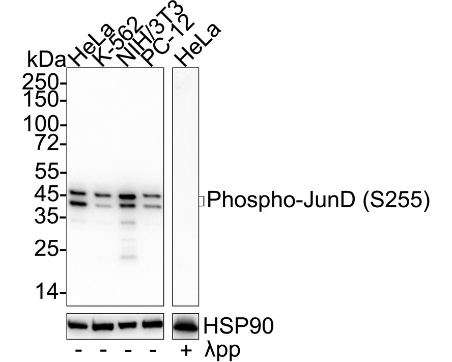
Western blot analysis of Phospho-JunD (S255) on different lysates with Rabbit anti-Phospho-JunD (S255) antibody at 1/5,000 dilution. Lane 1: HeLa cell lysate, Lane 2: K-562 cell lysate, Lane 3: NIH/3T3 cell lysate, Lane 4: PC-12 cell lysate, Lane 5: HeLa cell lysate, the membrane treated with λpp for 1 hour, Lysates/proteins at 20 µg/Lane. Exposure time: 30 seconds; 4-20% SDS-PAGE gel. Proteins were transferred to a PVDF membrane and blocked with 5% NFDM/TBST for 1 hour at room temperature. The primary antibody at 1/5,000 dilution was used in primary antibody dilution at 4℃ overnight. Goat Anti-Rabbit IgG - HRP Secondary Antibody at 1/50,000 dilution was used for 1 hour at room temperature.ICC/IF
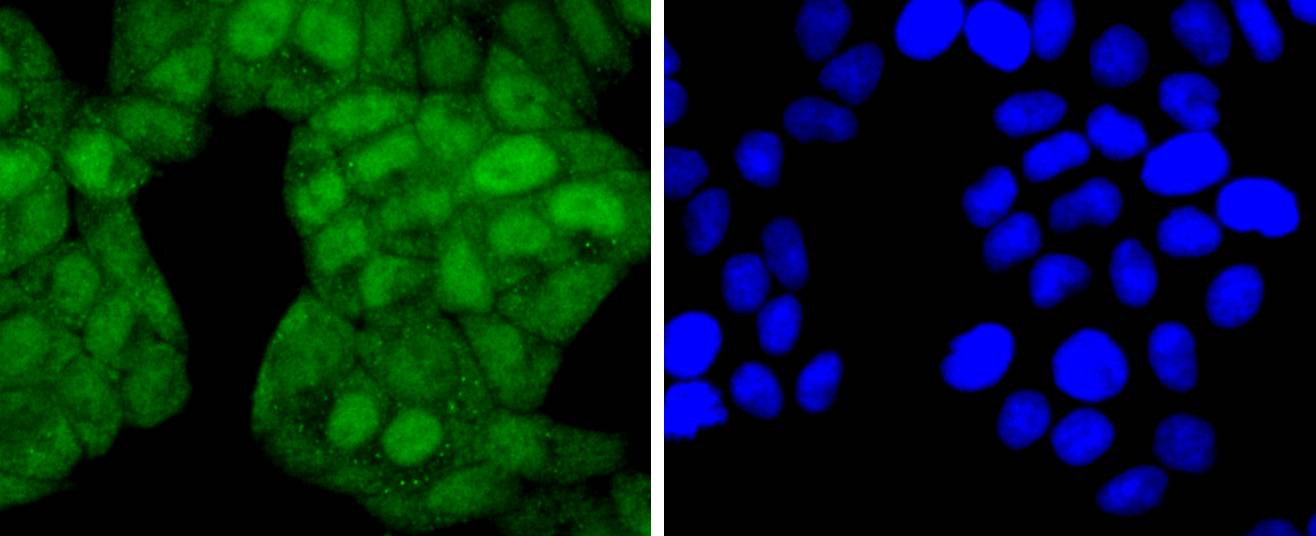
ICC staining Phospho-JunD(S255) in Hela cells (green). The nuclear counter stain is DAPI (blue). Cells were fixed in paraformaldehyde, permeabilised with 0.25% Triton X100/PBS.ICC/IF
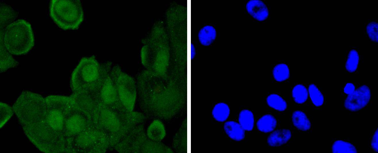
ICC staining Phospho-JunD(S255) in MCF-7 cells (green). The nuclear counter stain is DAPI (blue). Cells were fixed in paraformaldehyde, permeabilised with 0.25% Triton X100/PBS.IHC
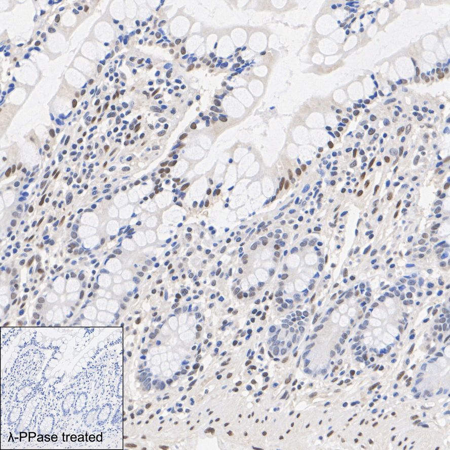
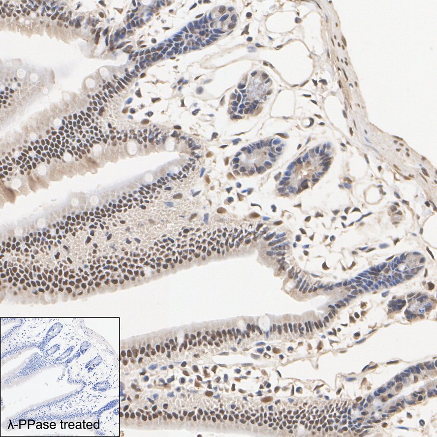
Immunohistochemical analysis of paraffin-embedded human small intestine tissue untreated / treated with λpp with Rabbit anti-Phospho-JunD (S255) antibody at 1/200 dilution. The section was pre-treated using heat mediated antigen retrieval with Tris-EDTA buffer (pH 9.0) for 20 minutes. The tissues were blocked in 1% BSA for 20 minutes at room temperature, washed with ddH2O and PBS, and then probed with the primary antibody at 1/200 dilution for 1 hour at room temperature. The detection was performed using an HRP conjugated compact polymer system. DAB was used as the chromogen. Tissues were counterstained with hematoxylin and mounted with DPX.IHC
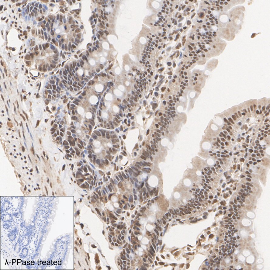
Immunohistochemical analysis of paraffin-embedded mouse small intestine tissue untreated / treated with λpp with Rabbit anti-Phospho-JunD (S255) antibody at 1/200 dilution. The section was pre-treated using heat mediated antigen retrieval with Tris-EDTA buffer (pH 9.0) for 20 minutes. The tissues were blocked in 1% BSA for 20 minutes at room temperature, washed with ddH2O and PBS, and then probed with the primary antibody at 1/200 dilution for 1 hour at room temperature. The detection was performed using an HRP conjugated compact polymer system. DAB was used as the chromogen. Tissues were counterstained with hematoxylin and mounted with DPX.IHC
Immunohistochemical analysis of paraffin-embedded rat small intestine tissue untreated / treated with λpp with Rabbit anti-Phospho-JunD (S255) antibody at 1/200 dilution. The section was pre-treated using heat mediated antigen retrieval with Tris-EDTA buffer (pH 9.0) for 20 minutes. The tissues were blocked in 1% BSA for 20 minutes at room temperature, washed with ddH2O and PBS, and then probed with the primary antibody at 1/200 dilution for 1 hour at room temperature. The detection was performed using an HRP conjugated compact polymer system. DAB was used as the chromogen. Tissues were counterstained with hematoxylin and mounted with DPX.| Product Name | Phospho-JunD (S255) Recombinant Rabbit Monoclonal Antibody |
|---|---|
| Antibody Type | Primary Antibodies |
| Product description | The activator protein-1 (AP-1) transcription factor consists of either Jun/Jun homodimers or Fos/Jun heterodimeric complexes. Homo- and heterodimers bind to the TGACTCA consensus sequence present in numerous promoters and initially identified as the phorbol ester tumor promoter response element (TRE). Jun B and Jun D have been shown to be almost identical to c-Jun in their C-terminal regions, which are involved in dimerization and DNA binding, whereas their N-terminal domains, which are involved in transcriptional activation, diverge. All three form heterodimers among themselves and with c-Fos and other members of the Fos gene family. Studies suggest that the two forms of Jun D may be due to internal initiation of translation. |
| Immunogen | Synthetic phospho-peptide corresponding to residues surrounding Ser255 of Human JunD aa 251-270 / 347. |
| Modification | p-S255 |
| Clonality | Monoclonal |
|---|---|
| Isotype | IgG |
| Host Species | Recombinant rabbit |
| Tested Applications | ICC/IFIHCWB |
| WB:1:5000 ICC/IF:1:50-1:200 IHC:1:50-1:200 |
|
| Species Reactivity | HumanMouseRat |
| Concentration | 1mg/ml |
| Purification | Protein A |
| Alternative Names | Activator protein 1 antibody AP 1 antibody AP1 antibody Jun D antibody jun D proto oncogene antibody Jund antibody JunD FL isoform antibody JUND_HUMAN antibody Transcription factor jun D antibody Transcription factor jun-D antibody |
|---|---|
| Molecular Weight(MW) | 35 kDa(Observed band size: 40/45 kDa) |
| Cellular Localization | Nucleus. |
WB
Western blot analysis of Phospho-JunD (S255) on different lysates with Rabbit anti-Phospho-JunD (S255) antibody at 1/5,000 dilution. Lane 1: HeLa cell lysate, Lane 2: K-562 cell lysate, Lane 3: NIH/3T3 cell lysate, Lane 4: PC-12 cell lysate, Lane 5: HeLa cell lysate, the membrane treated with λpp for 1 hour, Lysates/proteins at 20 µg/Lane. Exposure time: 30 seconds; 4-20% SDS-PAGE gel. Proteins were transferred to a PVDF membrane and blocked with 5% NFDM/TBST for 1 hour at room temperature. The primary antibody at 1/5,000 dilution was used in primary antibody dilution at 4℃ overnight. Goat Anti-Rabbit IgG - HRP Secondary Antibody at 1/50,000 dilution was used for 1 hour at room temperature.
ICC/IF
ICC staining Phospho-JunD(S255) in Hela cells (green). The nuclear counter stain is DAPI (blue). Cells were fixed in paraformaldehyde, permeabilised with 0.25% Triton X100/PBS.
ICC/IF
ICC staining Phospho-JunD(S255) in MCF-7 cells (green). The nuclear counter stain is DAPI (blue). Cells were fixed in paraformaldehyde, permeabilised with 0.25% Triton X100/PBS.
IHC
Immunohistochemical analysis of paraffin-embedded human small intestine tissue untreated / treated with λpp with Rabbit anti-Phospho-JunD (S255) antibody at 1/200 dilution. The section was pre-treated using heat mediated antigen retrieval with Tris-EDTA buffer (pH 9.0) for 20 minutes. The tissues were blocked in 1% BSA for 20 minutes at room temperature, washed with ddH2O and PBS, and then probed with the primary antibody at 1/200 dilution for 1 hour at room temperature. The detection was performed using an HRP conjugated compact polymer system. DAB was used as the chromogen. Tissues were counterstained with hematoxylin and mounted with DPX.
IHC
Immunohistochemical analysis of paraffin-embedded mouse small intestine tissue untreated / treated with λpp with Rabbit anti-Phospho-JunD (S255) antibody at 1/200 dilution. The section was pre-treated using heat mediated antigen retrieval with Tris-EDTA buffer (pH 9.0) for 20 minutes. The tissues were blocked in 1% BSA for 20 minutes at room temperature, washed with ddH2O and PBS, and then probed with the primary antibody at 1/200 dilution for 1 hour at room temperature. The detection was performed using an HRP conjugated compact polymer system. DAB was used as the chromogen. Tissues were counterstained with hematoxylin and mounted with DPX.
IHC
Immunohistochemical analysis of paraffin-embedded rat small intestine tissue untreated / treated with λpp with Rabbit anti-Phospho-JunD (S255) antibody at 1/200 dilution. The section was pre-treated using heat mediated antigen retrieval with Tris-EDTA buffer (pH 9.0) for 20 minutes. The tissues were blocked in 1% BSA for 20 minutes at room temperature, washed with ddH2O and PBS, and then probed with the primary antibody at 1/200 dilution for 1 hour at room temperature. The detection was performed using an HRP conjugated compact polymer system. DAB was used as the chromogen. Tissues were counterstained with hematoxylin and mounted with DPX.| Application Notes | WB:1:5000 ICC/IF:1:50-1:200 IHC:1:50-1:200 |
|---|
| Form | Liquid |
|---|---|
| Storage Instructions | Store at +4℃ after thawing. Aliquot store at -20℃ or -80℃. Avoid repeated freeze / thaw cycles. |
| Storage Buffer | 1*TBS (pH7.4), 1%BSA, 40%Glycerol. Preservative: 0.05% Sodium Azide. |


