WB
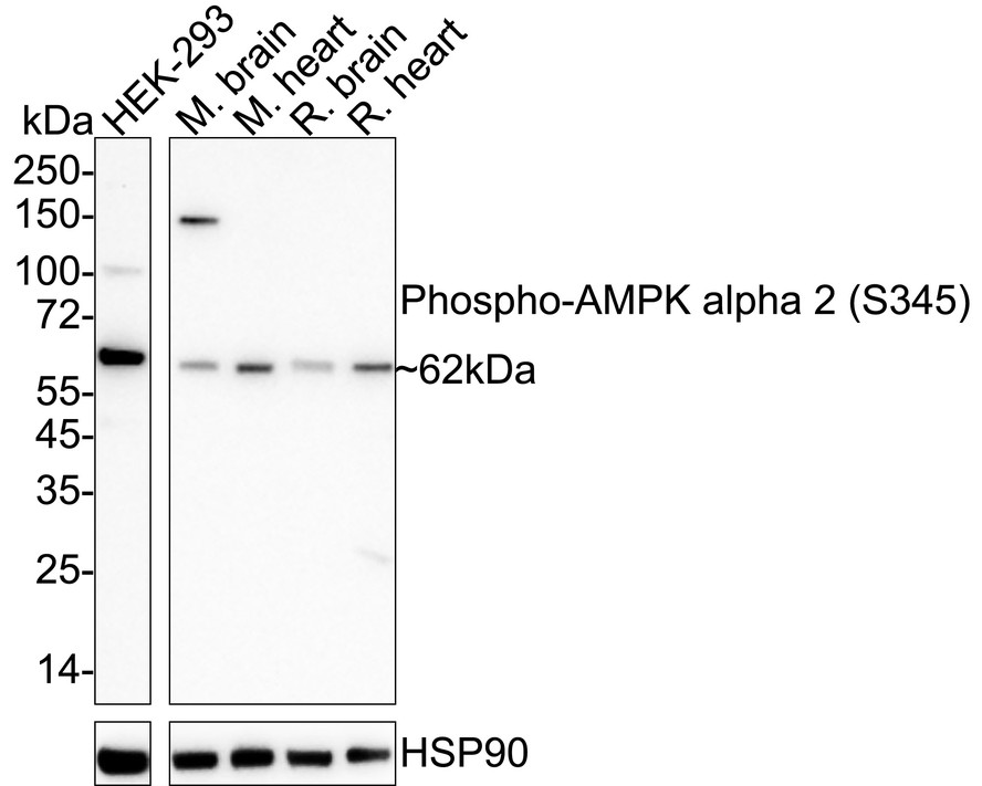
Western blot analysis of Phospho-AMPK alpha 2 (S345) on different lysates with Rabbit anti-Phospho-AMPK alpha 2 (S345) antibody at 1/1,000 dilution. Lane 1: HEK-293 cell lysate (20 µg/Lane), Lane 2: Mouse brain tissue lysate (40 µg/Lane), Lane 3: Mouse heart tissue lysate (40 µg/Lane), Lane 4: Rat brain tissue lysate (40 µg/Lane), Lane 5: Rat heart tissue lysate (40 µg/Lane), Exposure time: 3 minutes; 4-20% SDS-PAGE gel. Proteins were transferred to a PVDF membrane and blocked with 5% NFDM/TBST for 1 hour at room temperature. The primary antibody at 1/1,000 dilution was used in 5% NFDM/TBST at 4℃ overnight. Goat Anti-Rabbit IgG - HRP Secondary Antibody at 1/50,000 dilution was used for 1 hour at room temperature.IHC
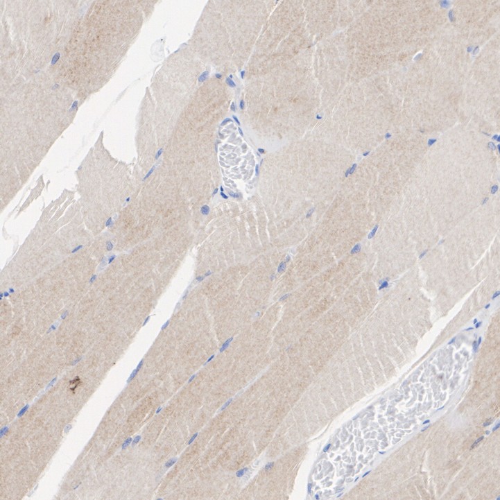
Immunohistochemical analysis of paraffin-embedded human skeletal muscle tissue with Rabbit anti-Phospho-AMPK alpha 2 (S345) antibody at 1/50 dilution. The section was pre-treated using heat mediated antigen retrieval with Tris-EDTA buffer (pH 9.0) for 20 minutes. The tissues were blocked in 1% BSA for 20 minutes at room temperature, washed with ddH2O and PBS, and then probed with the primary antibody at 1/50 dilution for 1 hour at room temperature. The detection was performed using an HRP conjugated compact polymer system. DAB was used as the chromogen. Tissues were counterstained with hematoxylin and mounted with DPX.IHC
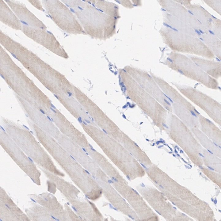
Immunohistochemical analysis of paraffin-embedded rat skeletal muscle tissue with Rabbit anti-Phospho-AMPK alpha 2 (S345) antibody at 1/50 dilution. The section was pre-treated using heat mediated antigen retrieval with Tris-EDTA buffer (pH 9.0) for 20 minutes. The tissues were blocked in 1% BSA for 20 minutes at room temperature, washed with ddH2O and PBS, and then probed with the primary antibody at 1/50 dilution for 1 hour at room temperature. The detection was performed using an HRP conjugated compact polymer system. DAB was used as the chromogen. Tissues were counterstained with hematoxylin and mounted with DPX.IHC
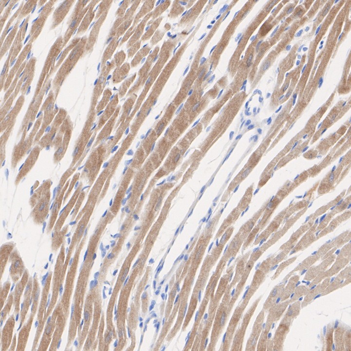
Immunohistochemical analysis of paraffin-embedded rat heart tissue with Rabbit anti-Phospho-AMPK alpha 2 (S345) antibody at 1/200 dilution. The section was pre-treated using heat mediated antigen retrieval with Tris-EDTA buffer (pH 9.0) for 20 minutes. The tissues were blocked in 1% BSA for 20 minutes at room temperature, washed with ddH2O and PBS, and then probed with the primary antibody at 1/50 dilution for 1 hour at room temperature. The detection was performed using an HRP conjugated compact polymer system. DAB was used as the chromogen. Tissues were counterstained with hematoxylin and mounted with DPX.FC
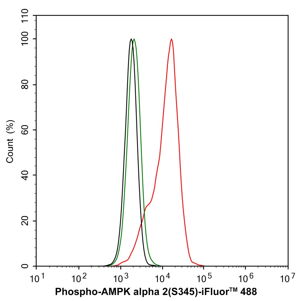
Flow cytometric analysis of HEK-293 cells labeling Phospho-AMPK alpha 2 (S345). Cells were fixed and permeabilized. Then stained with the primary antibody (1/1,000) (red) compared with Rabbit IgG Isotype Control (green). After incubation of the primary antibody at +4℃ for an hour, the cells were stained with a 488 conjugate-Goat anti-Rabbit IgG Secondary antibody at 1/1,000 dilution for 30 minutes at +4℃. Unlabelled sample was used as a control (cells without incubation with primary antibody; black).| Product Name | Phospho-AMPK alpha 2 (S345) Recombinant Rabbit Monoclonal Antibody |
|---|---|
| Antibody Type | Primary Antibodies |
| Product description | Five-prime-AMP-activated protein kinase, known as AMPK, is a heterotrimeric complex that comprises of a catalytic α subunit, and regulatory β and γ. AMPK protects cells from stresses that cause ATP depletion by switching off ATP-consuming biosynthetic pathways. AMPK is activated by high AMP and low ATP via a mechanism involving allosteric regulation, promotion of phosphorylation by an upstream protein kinase known as AMPK kinase (AMPKK), and inhibition of dephosphorylation. Activated AMPK can phosphorylate and regulate in vivo hydroxymethylglutaryl-CoA reductase and acetyl-CoA carboxylase, which are key regulatory enzymes of sterol synthesis and fatty acid synthesis, respectively. The human AMPKα1 gene maps to chromosome 5p12 and encodes a 548 amino acid protein. The major regulatory site phosphorylated by AMPKK on AMPKα has been identified as Thr 172 within the activation loop between the DFG and APE motifs of the alpha-subunits. |
| Immunogen | Synthetic phospho-peptide corresponding to residues surrounding Ser345 of Human AMPK alpha 2 aa 338-352 / 552. |
| Modification | p-S345 |
| Clonality | Monoclonal |
|---|---|
| Isotype | IgG |
| Host Species | Recombinant rabbit |
| Tested Applications | FCIHCWB |
| WB:1:500-1:2000 IHC:1:50-1:200 FC:1:1000 |
|
| Species Reactivity | HumanMouseRat |
| Concentration | 1mg/ml |
| Purification | Protein A |
| Alternative Names | 5'-AMP-activated protein kinase catalytic subunit alpha-2 antibody AAPK2_HUMAN antibody ACACA kinase antibody Acetyl-CoA carboxylase kinase antibody AMPK alpha 2 chain antibody AMPK subunit alpha-2 antibody AMPK2 antibody AMPKa2 antibody AMPKalpha2 antibody HMGCR kinase antibody Hydroxymethylglutaryl-CoA reductase kinase antibody PRKAA antibody PRKAA2 antibody Protein kinase AMP activated alpha 2 catalytic subunit antibody Protein kinase AMP activated catalytic subunit alpha 2 antibody |
|---|---|
| Molecular Weight(MW) | 62 kDa |
| Cellular Localization | Cytoplasm, Nucleus. |
WB
Western blot analysis of Phospho-AMPK alpha 2 (S345) on different lysates with Rabbit anti-Phospho-AMPK alpha 2 (S345) antibody at 1/1,000 dilution. Lane 1: HEK-293 cell lysate (20 µg/Lane), Lane 2: Mouse brain tissue lysate (40 µg/Lane), Lane 3: Mouse heart tissue lysate (40 µg/Lane), Lane 4: Rat brain tissue lysate (40 µg/Lane), Lane 5: Rat heart tissue lysate (40 µg/Lane), Exposure time: 3 minutes; 4-20% SDS-PAGE gel. Proteins were transferred to a PVDF membrane and blocked with 5% NFDM/TBST for 1 hour at room temperature. The primary antibody at 1/1,000 dilution was used in 5% NFDM/TBST at 4℃ overnight. Goat Anti-Rabbit IgG - HRP Secondary Antibody at 1/50,000 dilution was used for 1 hour at room temperature.
IHC
Immunohistochemical analysis of paraffin-embedded human skeletal muscle tissue with Rabbit anti-Phospho-AMPK alpha 2 (S345) antibody at 1/50 dilution. The section was pre-treated using heat mediated antigen retrieval with Tris-EDTA buffer (pH 9.0) for 20 minutes. The tissues were blocked in 1% BSA for 20 minutes at room temperature, washed with ddH2O and PBS, and then probed with the primary antibody at 1/50 dilution for 1 hour at room temperature. The detection was performed using an HRP conjugated compact polymer system. DAB was used as the chromogen. Tissues were counterstained with hematoxylin and mounted with DPX.
IHC
Immunohistochemical analysis of paraffin-embedded rat skeletal muscle tissue with Rabbit anti-Phospho-AMPK alpha 2 (S345) antibody at 1/50 dilution. The section was pre-treated using heat mediated antigen retrieval with Tris-EDTA buffer (pH 9.0) for 20 minutes. The tissues were blocked in 1% BSA for 20 minutes at room temperature, washed with ddH2O and PBS, and then probed with the primary antibody at 1/50 dilution for 1 hour at room temperature. The detection was performed using an HRP conjugated compact polymer system. DAB was used as the chromogen. Tissues were counterstained with hematoxylin and mounted with DPX.
IHC
Immunohistochemical analysis of paraffin-embedded rat heart tissue with Rabbit anti-Phospho-AMPK alpha 2 (S345) antibody at 1/200 dilution. The section was pre-treated using heat mediated antigen retrieval with Tris-EDTA buffer (pH 9.0) for 20 minutes. The tissues were blocked in 1% BSA for 20 minutes at room temperature, washed with ddH2O and PBS, and then probed with the primary antibody at 1/50 dilution for 1 hour at room temperature. The detection was performed using an HRP conjugated compact polymer system. DAB was used as the chromogen. Tissues were counterstained with hematoxylin and mounted with DPX.
FC
Flow cytometric analysis of HEK-293 cells labeling Phospho-AMPK alpha 2 (S345). Cells were fixed and permeabilized. Then stained with the primary antibody (1/1,000) (red) compared with Rabbit IgG Isotype Control (green). After incubation of the primary antibody at +4℃ for an hour, the cells were stained with a 488 conjugate-Goat anti-Rabbit IgG Secondary antibody at 1/1,000 dilution for 30 minutes at +4℃. Unlabelled sample was used as a control (cells without incubation with primary antibody; black).| Application Notes | WB:1:500-1:2000 IHC:1:50-1:200 FC:1:1000 |
|---|
| Form | Liquid |
|---|---|
| Storage Instructions | Store at +4℃ after thawing. Aliquot store at -20℃ or -80℃. Avoid repeated freeze / thaw cycles. |
| Storage Buffer | 1*TBS (pH7.4), 1%BSA, 40%Glycerol. Preservative: 0.05% Sodium Azide. |


