Application
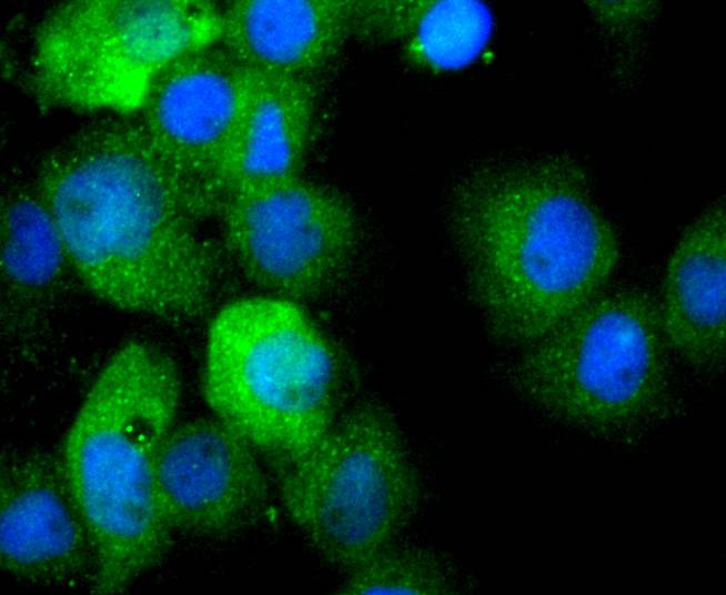
Fig1: ICC staining IL1 beta in A431 cells (green). The nuclear counter stain is DAPI (blue). Cells were fixed in paraformaldehyde, permeabilised with 0.25% Triton X100/PBS.Application
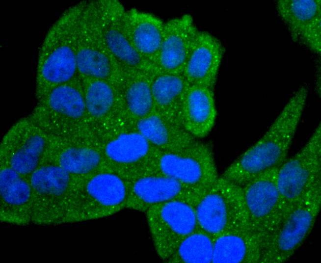
Fig2: ICC staining IL1 beta in Hela cells (green). The nuclear counter stain is DAPI (blue). Cells were fixed in paraformaldehyde, permeabilised with 0.25% Triton X100/PBS.Application
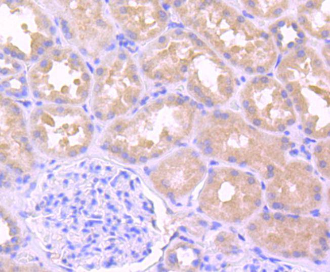
Fig3: Immunohistochemical analysis of paraffin-embedded human kidney tissue using anti-IL1 beta antibody. Counter stained with hematoxylin.Application
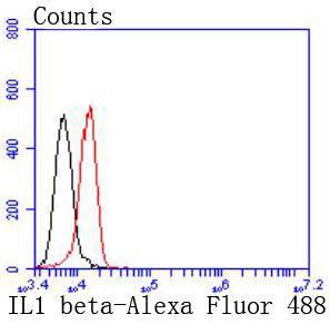
Fig4: Flow cytometric analysis of Hela cells with IL1 beta antibody at 1/50 dilution (red) compared with an unlabelled control (cells without incubation with primary antibody; black). Alexa Fluor 488-conjugated goat anti rabbit IgG was used as the secondary antibody| Product Name | IL1 beta [JJ087-3] |
|---|---|
| Antibody Type | Primary Antibodies |
| Antigen Alias | Catabolin antibody H1 antibody IL 1 antibody IL 1 beta antibody IL-1 beta antibody IL1 BETA antibody IL1B antibody IL1B_HUMAN antibody IL1F2 antibody Interleukin 1 beta antibody Interleukin-1 beta antibody OAF antibody OTTHUMP00000162031 antibody Preinterleukin 1 beta antibody Pro interleukin 1 beta antibody |
| Product description | Two forms of interleukin-1, designated IL-1α and IL-1β, have been described. Although encoded by distinct genes and exhibiting roughly only 25% sequence identity, IL-1α and IL-1β bind to the same receptor and seem to elicit similar biological responses. IL-1 production is generally thought to be associated with inflammation, but it has also been shown to be expressed during kidney development, thymocyte differentiation and cartilage degradation. IL-1 plays a critical role in the regulation of immune response and inflammation, acting as an activator of T and B lymphocytes and natural killer (NK) cells. In T cells, IL-1 stimulates the production of IL-2 and selectively inhibits IL-4 expression. IL-1 induces B cell proliferation and maturation, and immunoglobulin synthesis. NK cells require IL-1β for production of the anti- pathogen IFN-γ. IL-1 has also been implicated in several pathological conditions including rheumatoid arthritis, inflammatory bowel disease and atherosclerosis. |
| Immunogen | recombinant protein |
| Clonality | Monoclonal |
|---|---|
| Isotype | IgG |
| Host Species | Recombinant rabbit |
| Tested Applications | WBICC/IFIHCFC |
| WB:1:1,000 ICC:1:50-1:200 IHC:1:50-1:200 FC:1:50-1:100 |
|
| Species Reactivity | Human |
| Concentration | 1mg/ml |
| Alternative Names | Catabolin antibody H1 antibody IL 1 antibody IL 1 beta antibody IL-1 beta antibody IL1 BETA antibody IL1B antibody IL1B_HUMAN antibody IL1F2 antibody Interleukin 1 beta antibody Interleukin-1 beta antibody OAF antibody OTTHUMP00000162031 antibody Preinterleukin 1 beta antibody Pro interleukin 1 beta antibody |
|---|---|
| Molecular Weight(MW) | 31 kDa |
| Cellular Localization | Cytoplasm, Lysosome, Secreted. |
| SwissProt ID | P01584 |
|---|
Application
Fig1: ICC staining IL1 beta in A431 cells (green). The nuclear counter stain is DAPI (blue). Cells were fixed in paraformaldehyde, permeabilised with 0.25% Triton X100/PBS.
Application
Fig2: ICC staining IL1 beta in Hela cells (green). The nuclear counter stain is DAPI (blue). Cells were fixed in paraformaldehyde, permeabilised with 0.25% Triton X100/PBS.
Application
Fig3: Immunohistochemical analysis of paraffin-embedded human kidney tissue using anti-IL1 beta antibody. Counter stained with hematoxylin.
Application
Fig4: Flow cytometric analysis of Hela cells with IL1 beta antibody at 1/50 dilution (red) compared with an unlabelled control (cells without incubation with primary antibody; black). Alexa Fluor 488-conjugated goat anti rabbit IgG was used as the secondary antibody| Positive Control | A431, Hela, human kidney tissue. |
|---|---|
| Application Notes | WB:1:1,000 ICC:1:50-1:200 IHC:1:50-1:200 FC:1:50-1:100 |
| Form | Liquid |
|---|---|
| Storage Instructions | Store at +4℃ after thawing. Aliquot store at -20℃ or -80℃. Avoid repeated freeze / thaw cycles. |
| Storage Buffer | 1*TBS (pH7.4), 1%BSA, 40%Glycerol. Preservative: 0.05% Sodium Azide. |


