Application
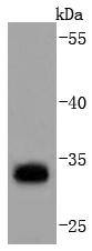
Fig1: Western blot analysis of PD-L1 on mouse heart lysates using anti-PD-L1 antibody at 1/1,000 dilution.Application
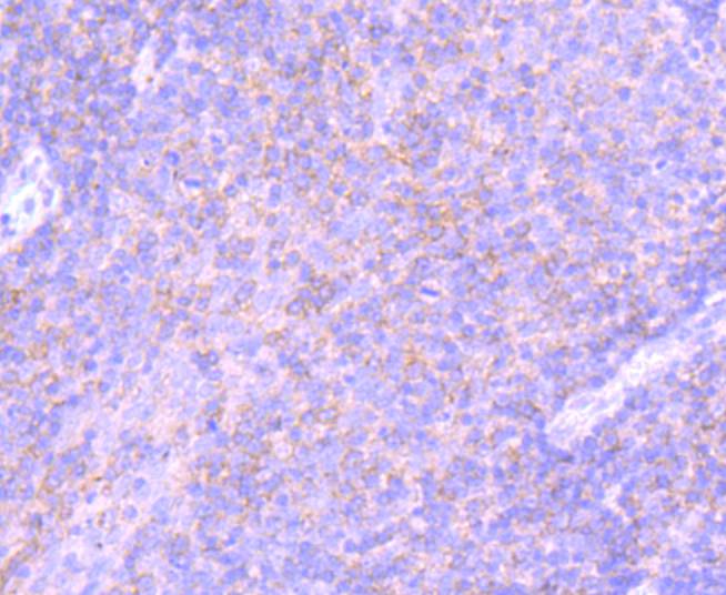
Fig2: Immunohistochemical analysis of paraffin-embedded human tonsil tissue using anti-PD-L1 antibody. Counter stained with hematoxylin.Application
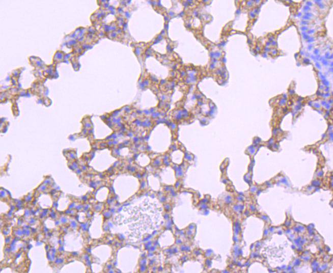
Fig3: Immunohistochemical analysis of paraffin-embedded mouse lung tissue using anti-PD-L1 antibody. Counter stained with hematoxylin.| Product Name | PD-L1 [JJ08-95] |
|---|---|
| Antibody Type | Primary Antibodies |
| Antigen Alias | B7 H antibody B7 H1 antibody B7 homolog 1 antibody B7-H1 antibody B7H antibody B7H1 antibody CD 274 antibody CD274 antibody CD274 antigen antibody CD274 molecule antibody MGC142294 antibody MGC142296 antibody OTTHUMP00000021029 antibody PD L1 antibody PD-L1 antibody PD1L1_HUMAN antibody PDCD1 ligand 1 antibody PDCD1L1 antibody PDCD1LG1 antibody PDL 1 antibody PDL1 antibody Programmed cell death 1 ligand 1 antibody Programmed death ligand 1 antibody RGD1566211 antibody |
| Product description | Engagement of CD28 by B7-1 (CD80) or B7-2 (CD86) in the presence of antigen promotes T cell proliferation, cytokine production, differentiation of effector T cells, and the induction of Bcl-x, a promoter of T cell survival. Conversely, engagement of CTLA4 by B7-1 or B7-2 may inhibit proliferation and IL-2 production. Pdcd-1L1 (programmed cell death ligand-1), also known as B7-H1 or PD-L1, is 290 amino acid type I transmembrane protein which is 20% and 15% identical to B7-1 and B7-2, respectively. Pdcd-1L2 has immunoglobulin V-like and C-like domains and a 30 amino acid cytoplasmic tail. It does not bind CD28, cytotoxic T-lymphocyte A4 or ICOS (inducible co-stimulator). IL-2, although produced in small amounts, is required for the effect of Pdcd-1L1 co-stimulation. The gene which encodes Pdcd-1L1 maps to human chromosome 9p24. Pdcd-1L2 (programmed cell death ligand-2) is a 73 amino acid protein which contains a signal sequence, IgV- and IgC-like domains, a transmembrane region and a cytoplasmic region. The gene which encodes Pdcd-1L2 maps to human chromosome 9p24.2. The constitutive expression of Pdcd-1L1 and Pdcd-1L2 on paren-chymal cells of heart, lung and kidney suggests that the Pdcd-1-Pdcd-L system could provide unique negative signaling to help prevent autoimmune disease. |
| Immunogen | recombinant protein |
| Clonality | Monoclonal |
|---|---|
| Isotype | IgG |
| Host Species | Recombinant rabbit |
| Tested Applications | WBIHC |
| WB:1:1,000 IHC:1:50-1:200 |
|
| Species Reactivity | HumanMouseRat |
| Concentration | 1mg/ml |
| Alternative Names | B7 H antibody B7 H1 antibody B7 homolog 1 antibody B7-H1 antibody B7H antibody B7H1 antibody CD 274 antibody CD274 antibody CD274 antigen antibody CD274 molecule antibody MGC142294 antibody MGC142296 antibody OTTHUMP00000021029 antibody PD L1 antibody PD-L1 antibody PD1L1_HUMAN antibody PDCD1 ligand 1 antibody PDCD1L1 antibody PDCD1LG1 antibody PDL 1 antibody PDL1 antibody Programmed cell death 1 ligand 1 antibody Programmed death ligand 1 antibody RGD1566211 antibody |
|---|---|
| Molecular Weight(MW) | 33 kDa |
| Cellular Localization | Cell membrane, Endomembrane system. |
Application
Fig1: Western blot analysis of PD-L1 on mouse heart lysates using anti-PD-L1 antibody at 1/1,000 dilution.
Application
Fig2: Immunohistochemical analysis of paraffin-embedded human tonsil tissue using anti-PD-L1 antibody. Counter stained with hematoxylin.
Application
Fig3: Immunohistochemical analysis of paraffin-embedded mouse lung tissue using anti-PD-L1 antibody. Counter stained with hematoxylin.| Positive Control | Human tonsil tissue, mouse heart tissue, human melanoma tissue, human lung cancer tissue. |
|---|---|
| Application Notes | WB:1:1,000 IHC:1:50-1:200 |
| Form | Liquid |
|---|---|
| Storage Instructions | Store at +4℃ after thawing. Aliquot store at -20℃ or -80℃. Avoid repeated freeze / thaw cycles. |
| Storage Buffer | 1*TBS (pH7.4), 1%BSA, 40%Glycerol. Preservative: 0.05% Sodium Azide. |


